,Dermatology – USMLE Step 2 CK 2025 – High-Yield
Clinical Review for Board Preparation
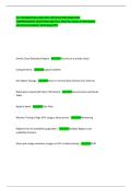
Non-bullous Impetigo
staph aureus or A beta-hemolytic strep
Vesicles → rupture → honey-colored crusts
Hepers Zoster syndromes
Ophthalmic branch: Lesion on tip of the nose (Hutchinson's sign)
Ramsey-Hunt:
o Vesicles on tympanic membrane, external auditory canal may be involved
o Loss of taste anterior 2/3 tongue unilaterally
o Bell's palsy
Trigeminal Nerve: forehead, perioccular region, nose
Varicella (Chicken Pox)
"Dewdrop on rose petal"
Lesions of different stages present at the same time: papules, vesicles, pustules, and crusts
Prodrome: low fever, headache, malaise directly before
intense pruritis
Toxic Epidermal Necrolysis
Affects 30-100% of skin surface
Targetoid lesions/bullae w/ sudden onset
Full-thickness epidermal detachment
+Nikolsky
Pemphigous Vulgaris
Widespread erosions where blisters have ruptured
+Nikolsky sign
oral erosions usually precede skin blisters
Dermatitis Herpetiformis
Associated w/ gluten sensitivity
Isolated or grouped, intensely burning/pruritic urticarial papules, vesicles and rarely bullae -->
erosions/crusts
Symmetrically distributed
, Erythema Nodosum
Nodules/plaques, erythematous, tender eruption → evolve into purple, red-brown (bruise-like look).
Joint pain
Mostly lower legs
commonly after HSV
Atopic Dermatitis
red, crusted, excoriated and lichenified patches and plaques
Asteatotic Eczema
eczema craquele
Eczema Herpeticum
Widespread HSV w/ atopic dermatitis/eczema; numerous umbilicated vesicles; may have fever
clinical syndrome of HSV
Benign melanocytic nevi: Junctional nevi
Uniform tan or brown macule - oval or round, smooth regular borders
Benign melanocytic nevi: Compound nevi
Tan or brown, smooth, sharply defined, round, soft papules (slightly or very raise)
some w/ coarse hairs
Benign melanocytic nevi: Intradermal nevi
Flesh-colored, tan or brown papule
Soft, dome-shaped
Some hairy
Congenital Melanocytic nevi
Tan to dark brown, macule or papule w/ irregular verrucous surface and terminal hair
Giant congenital nevi - garment distribution
Congenital Melanocytic nevi
Tan to dark brown, macule or papule w/ irregular verrucous surface and terminal hair
Giant congenital nevi - garment distribution
Superficial Spreading Melanoma
Most common type
Red, white, blue, brown, black paupule or nodule
Associated with precursor melanocytic nevi
Nodular Melanomas
Clinical Review for Board Preparation
Non-bullous Impetigo
staph aureus or A beta-hemolytic strep
Vesicles → rupture → honey-colored crusts
Hepers Zoster syndromes
Ophthalmic branch: Lesion on tip of the nose (Hutchinson's sign)
Ramsey-Hunt:
o Vesicles on tympanic membrane, external auditory canal may be involved
o Loss of taste anterior 2/3 tongue unilaterally
o Bell's palsy
Trigeminal Nerve: forehead, perioccular region, nose
Varicella (Chicken Pox)
"Dewdrop on rose petal"
Lesions of different stages present at the same time: papules, vesicles, pustules, and crusts
Prodrome: low fever, headache, malaise directly before
intense pruritis
Toxic Epidermal Necrolysis
Affects 30-100% of skin surface
Targetoid lesions/bullae w/ sudden onset
Full-thickness epidermal detachment
+Nikolsky
Pemphigous Vulgaris
Widespread erosions where blisters have ruptured
+Nikolsky sign
oral erosions usually precede skin blisters
Dermatitis Herpetiformis
Associated w/ gluten sensitivity
Isolated or grouped, intensely burning/pruritic urticarial papules, vesicles and rarely bullae -->
erosions/crusts
Symmetrically distributed
, Erythema Nodosum
Nodules/plaques, erythematous, tender eruption → evolve into purple, red-brown (bruise-like look).
Joint pain
Mostly lower legs
commonly after HSV
Atopic Dermatitis
red, crusted, excoriated and lichenified patches and plaques
Asteatotic Eczema
eczema craquele
Eczema Herpeticum
Widespread HSV w/ atopic dermatitis/eczema; numerous umbilicated vesicles; may have fever
clinical syndrome of HSV
Benign melanocytic nevi: Junctional nevi
Uniform tan or brown macule - oval or round, smooth regular borders
Benign melanocytic nevi: Compound nevi
Tan or brown, smooth, sharply defined, round, soft papules (slightly or very raise)
some w/ coarse hairs
Benign melanocytic nevi: Intradermal nevi
Flesh-colored, tan or brown papule
Soft, dome-shaped
Some hairy
Congenital Melanocytic nevi
Tan to dark brown, macule or papule w/ irregular verrucous surface and terminal hair
Giant congenital nevi - garment distribution
Congenital Melanocytic nevi
Tan to dark brown, macule or papule w/ irregular verrucous surface and terminal hair
Giant congenital nevi - garment distribution
Superficial Spreading Melanoma
Most common type
Red, white, blue, brown, black paupule or nodule
Associated with precursor melanocytic nevi
Nodular Melanomas