Chapter 15 – Digestive System Development
Overview
• The digestive system is derived from:
o Endoderm: Forms the epithelial lining of the gut tube and parenchyma of glands (e.g.,
hepatocytes, pancreas).
o Mesoderm: Gives rise to muscle, connective tissue, and peritoneal components.
• Primitive Gut Tube Formation:
o Results from cephalocaudal and lateral folding of the embryo.
o Formed as part of the endoderm-lined yolk sac cavity is incorporated into the
embryo.
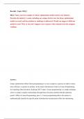
• Gut Tube Regions:
1. Pharyngeal gut: From oropharyngeal membrane to the respiratory diverticulum.
2. Foregut: From the pharynx to the liver outgrowth.
3. Midgut: From caudal to the liver bud to the right 2/3 of the transverse colon.
4. Hindgut: From the left 1/3 of the transverse colon to the cloacal membrane.
Development of the Gut Tube
Gut Tube Layers:
• Endoderm:
o Forms the epithelial lining and glandular parenchyma.
• Visceral Mesoderm:
o Gives rise to connective tissue, smooth muscle, and blood vessels.
Molecular Regulation
• Retinoic Acid (RA) Gradient:
o Low RA: Pharynx.
o High RA: Colon.
o Drives expression of region-specific transcription factors:
§ SOX2: Esophagus and stomach.
§ PDX1: Duodenum.
§ CDXC: Small intestine.
§ CDXA: Large intestine and rectum.
• Sonic Hedgehog (SHH):
o Secreted by endodermal cells.
o Activates mesodermal HOX genes, determining the type of structure formed.
Mesenteries
• Gut tube is suspended by mesenteries, which are double layers of peritoneum.
, o Dorsal Mesentery:
§ Present along the entire length of the gut tube.
§ Forms:
§ Dorsal mesogastrium: Stomach region.
§ Dorsal mesoduodenum: Duodenum region.
§ Dorsal mesocolon: Colon region.
§ Mesentery proper: Jejunal and ileal loops.
o Ventral Mesentery:
§ Limited to foregut derivatives.
§ Derived from the septum transversum.
§ Forms:
§ Falciform ligament: Liver to ventral body wall.
§ Lesser omentum: Liver to stomach and duodenum.
Foregut Development
Esophagus
• 4th Week: Forms from the caudal foregut.
• Tracheoesophageal septum divides foregut into:
o Posterior esophagus.
o Anterior trachea and lung buds.
• Muscular Development:
o Upper 1/3: Skeletal muscle from pharyngeal arch mesenchyme.
o Lower 2/3: Smooth muscle from splanchnic mesoderm.
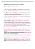
Stomach
• Appears as a dilation of the foregut in the 4th week.
• Rotations:
1. Longitudinal Axis Rotation:
§ Rotates 90° clockwise.
§ Left side becomes anterior; right side becomes posterior.
2. Anteroposterior Axis Rotation:
§ Pyloric region moves upward and right.
§ Cardiac region moves downward and left.
• Unequal growth forms:
o Greater curvature: Posterior wall grows faster.
o Lesser curvature: Anterior wall grows slower.
• Associated Structures:
Overview
• The digestive system is derived from:
o Endoderm: Forms the epithelial lining of the gut tube and parenchyma of glands (e.g.,
hepatocytes, pancreas).
o Mesoderm: Gives rise to muscle, connective tissue, and peritoneal components.
• Primitive Gut Tube Formation:
o Results from cephalocaudal and lateral folding of the embryo.
o Formed as part of the endoderm-lined yolk sac cavity is incorporated into the
embryo.
• Gut Tube Regions:
1. Pharyngeal gut: From oropharyngeal membrane to the respiratory diverticulum.
2. Foregut: From the pharynx to the liver outgrowth.
3. Midgut: From caudal to the liver bud to the right 2/3 of the transverse colon.
4. Hindgut: From the left 1/3 of the transverse colon to the cloacal membrane.
Development of the Gut Tube
Gut Tube Layers:
• Endoderm:
o Forms the epithelial lining and glandular parenchyma.
• Visceral Mesoderm:
o Gives rise to connective tissue, smooth muscle, and blood vessels.
Molecular Regulation
• Retinoic Acid (RA) Gradient:
o Low RA: Pharynx.
o High RA: Colon.
o Drives expression of region-specific transcription factors:
§ SOX2: Esophagus and stomach.
§ PDX1: Duodenum.
§ CDXC: Small intestine.
§ CDXA: Large intestine and rectum.
• Sonic Hedgehog (SHH):
o Secreted by endodermal cells.
o Activates mesodermal HOX genes, determining the type of structure formed.
Mesenteries
• Gut tube is suspended by mesenteries, which are double layers of peritoneum.
, o Dorsal Mesentery:
§ Present along the entire length of the gut tube.
§ Forms:
§ Dorsal mesogastrium: Stomach region.
§ Dorsal mesoduodenum: Duodenum region.
§ Dorsal mesocolon: Colon region.
§ Mesentery proper: Jejunal and ileal loops.
o Ventral Mesentery:
§ Limited to foregut derivatives.
§ Derived from the septum transversum.
§ Forms:
§ Falciform ligament: Liver to ventral body wall.
§ Lesser omentum: Liver to stomach and duodenum.
Foregut Development
Esophagus
• 4th Week: Forms from the caudal foregut.
• Tracheoesophageal septum divides foregut into:
o Posterior esophagus.
o Anterior trachea and lung buds.
• Muscular Development:
o Upper 1/3: Skeletal muscle from pharyngeal arch mesenchyme.
o Lower 2/3: Smooth muscle from splanchnic mesoderm.
Stomach
• Appears as a dilation of the foregut in the 4th week.
• Rotations:
1. Longitudinal Axis Rotation:
§ Rotates 90° clockwise.
§ Left side becomes anterior; right side becomes posterior.
2. Anteroposterior Axis Rotation:
§ Pyloric region moves upward and right.
§ Cardiac region moves downward and left.
• Unequal growth forms:
o Greater curvature: Posterior wall grows faster.
o Lesser curvature: Anterior wall grows slower.
• Associated Structures: