Xzone SPI Exam Questions And Answers
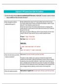
This is a color Doppler of a normal artery and vein. What would a
sonographer adjust in order to change the appearance of the upper
vessel to a single uniform color?
- use harmonics
- increase the wall filter
- increase the scale
- increase the overall gain
- decrease the wall filter -
correct answer ✅increase the scale
The two circles in the image below are a normal artery and a vein.
What adjustment on the system would you make to fill the lumen
of vessel #2 with color?
- increase the scale
,Xzone SPI Exam Questions And Answers
- increase the color gain
- decrease the scale
- increase the overall gain
- increase harmonics -
correct answer ✅decrease the scale
Which of the following statements best describes the Doppler
spectrum below?
- Low velocity flow does not appear because the Doppler gain
setting is incorrect
- Low velocity flow does not appear because the sample volume is
not positioned in an area with color in the gray scale image
- Low amplitude flow does not appear because the wall filter is set
at an incorrect level
,Xzone SPI Exam Questions And Answers
- Low velocity flow does not appear because the wall filter is set at
an incorrect level
- Low amplitude flow does not appear because the overall gain
setting is incorrect -
correct answer ✅Low velocity flow does not appear because the
wall filter is set at an incorrect level
What is direction of flow of blood in the vessel appearing below?
- From the right of the screen to the left of the screen.
- Away from the transducer
- From the left of the screen to the right of the screen.
- Toward the transducer. -
correct answer ✅From the right of the screen to the left of the
screen.
What is going on with this Doppler image?
- This is an image created with a combination of CT and ultrasound.
- This is an image created with power mode Doppler.
, Xzone SPI Exam Questions And Answers
- This image results from all the flow moving away from the
transducer.
- Who knows what is going on?
- Unidirectional color Doppler created this image -
correct answer ✅This is an image created with power mode
Doppler.
Which of the following steps can the sonographer perform to
create color within the vessel in this image?
- Use a wall filter.
- Use angle correction.
- Steer the Doppler box straight down.
- Increase the color Doppler gain.