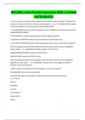
BIO-202L Latest Practice Questions 2025 | Verified
and Graded A+
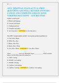
Fill-in the sentences with the correct organ that match the function described. Then place the
sentences in order of the GI tract, from proximal to distal. - 1. The ORAL CAVITY provides
mechanical digestion of chewing, which is called MASTICATION.
2. The ESOPHAGUS moves food from the pharynx to the STOMACH with wave like contractions
called PERISTALTIC WAVES.
3. The STOMACH is a hollow storage vat that initiates digest of proteins.
4. The SMALL INTESTINES is where most absorption of nutrients takes place.
5. The LARGE INTESTINES absorbs a little water and electroytes, but is mostly for storing feces.
Place the appropriate words and descriptions with the picture with the correct highlighted
salivary gland. - SUBLINGUAL GLAND: produces 3-5% of saliva
SUBMANDIBULAR GLAND: produces 60-70% of saliva
PAROTID GLAND: produces 25-30% of saliva
Place the appropriate words and descriptions with the picture with the correct highlighted
digestive accessory organ. - PANCREAS: Produces and secretes digestive enzymes.
Releases insulin into the blood.
LIVER: Produces and secretes bile. Stores glycogen, minerals and vitamins.
GALLBLADDER: Stores and concentrate bile.
Label ONLY the organs of the digestive system that comprise the alimentary tract. -
mouth
pharynx
esophagus
stomach
small intestine
large intestine
rectum
,anus
Label ONLY the accessory organs of the digestive system. - liver
gallbladder
salivary glands
pancreas
Drag each label into the appropriate box based on its role in the digestive system. -
ACCESSORY ORGANS:
teeth
salivary glands
pancreas
liver
gallbladder
DIGESTIVE TRACT:
oral cavity
stomach
esophagus
small intestine
large intestine
Place the following anatomical structures in the correct order, following the path that food
would take. - 1. oral cavity
2. pharynx
3. esophagus
4. stomach
5. small intestine
6. large intestine
, 7. rectum
8. anus
Place the following anatomical structures in the correct order, following the path that food
would take, starting with the stomach. - 1. oral cavity
2. pharynx
3. esophagus
4. stomach
5. duodenum
6. jejunum
7. ileum
The figure shows a portion of the wall of the small intestine. Label the layers of the digestive
tract wall and the associated structures. - lacteal
villi
intestinal gland
nerve plexuses
longitudinal muscle
serosa
muscular layer
submucosa
mucosa
Label the structures associated with the mucosa of the digestive tract wall. - microvilli
goblet cel
nucleus
simple columnar epithelium
capillary
lacteal
and Graded A+
Fill-in the sentences with the correct organ that match the function described. Then place the
sentences in order of the GI tract, from proximal to distal. - 1. The ORAL CAVITY provides
mechanical digestion of chewing, which is called MASTICATION.
2. The ESOPHAGUS moves food from the pharynx to the STOMACH with wave like contractions
called PERISTALTIC WAVES.
3. The STOMACH is a hollow storage vat that initiates digest of proteins.
4. The SMALL INTESTINES is where most absorption of nutrients takes place.
5. The LARGE INTESTINES absorbs a little water and electroytes, but is mostly for storing feces.
Place the appropriate words and descriptions with the picture with the correct highlighted
salivary gland. - SUBLINGUAL GLAND: produces 3-5% of saliva
SUBMANDIBULAR GLAND: produces 60-70% of saliva
PAROTID GLAND: produces 25-30% of saliva
Place the appropriate words and descriptions with the picture with the correct highlighted
digestive accessory organ. - PANCREAS: Produces and secretes digestive enzymes.
Releases insulin into the blood.
LIVER: Produces and secretes bile. Stores glycogen, minerals and vitamins.
GALLBLADDER: Stores and concentrate bile.
Label ONLY the organs of the digestive system that comprise the alimentary tract. -
mouth
pharynx
esophagus
stomach
small intestine
large intestine
rectum
,anus
Label ONLY the accessory organs of the digestive system. - liver
gallbladder
salivary glands
pancreas
Drag each label into the appropriate box based on its role in the digestive system. -
ACCESSORY ORGANS:
teeth
salivary glands
pancreas
liver
gallbladder
DIGESTIVE TRACT:
oral cavity
stomach
esophagus
small intestine
large intestine
Place the following anatomical structures in the correct order, following the path that food
would take. - 1. oral cavity
2. pharynx
3. esophagus
4. stomach
5. small intestine
6. large intestine
, 7. rectum
8. anus
Place the following anatomical structures in the correct order, following the path that food
would take, starting with the stomach. - 1. oral cavity
2. pharynx
3. esophagus
4. stomach
5. duodenum
6. jejunum
7. ileum
The figure shows a portion of the wall of the small intestine. Label the layers of the digestive
tract wall and the associated structures. - lacteal
villi
intestinal gland
nerve plexuses
longitudinal muscle
serosa
muscular layer
submucosa
mucosa
Label the structures associated with the mucosa of the digestive tract wall. - microvilli
goblet cel
nucleus
simple columnar epithelium
capillary
lacteal