CASE 1: Blood cells, coagulation & inflammtion
zondag 15 oktober 2023 11:11
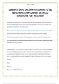
1. Types of blood cells, structure, function, and origin
It starts with a hematopoietic stem cell. This then splits into the lymphoid group
and the myeloid group.
The Myeloid group
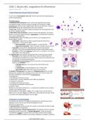
1. Red blood cells/ Enterocytes: lack a nucleus and organelles and contain
haemoglobin (oxygen transport and gas exchange). The production of RBCs
Erythropoiesis is driven by low oxygen levels in the tissues and occurs in the red
bone marrow (where the hematopoietic stem cell is present)
2. Mast cells: mediate host defence against pathogens by releasing granules and
are active in allergic reactions
3. White blood cells/ Leukocytes: contain a nucleus and organelles. The overall
function is protection against diseases. Leukopoiesis is stimulated by interleukins.
They can be subdivided into
3.1 Granulocytes: bigger than RBCs, but live shorter, are all phagocytes to a
certain degree. Contain:
- Neutrophils: responsible for defence against bacterial infections by
phagocytosis. (most present WBC)
○ Band neutrophils = young neutrophils, 1 nucleus particle
○ Segmented neutrophils = older neutrophil, multiple nucleus particles.
- Eosinophils: responsible for fighting allergic reactions and parasitic
infections. They can be recognized by the red colour and bi-lobed nucleus
- Basophils: are filled with Histamine, which acts as a vasodilator and attracts
other WBC to the inflamed site.
3.2 Agranulocytes: lack cytoplasmatic granules their nuclei are kidney shaped.
- Monocytes are the largest cell in the blood , they can differentiate into
macrophages and protect from viruses, bacteria and chronic infections. And
are important in activating lymphocytes.
- Lymphocytes (lymphoid group): play a crucial role in immunity by direct cell
attach or via antibodies
○ B-lymphocytes: give rise to plasma cells which will produce
antibodies.
○ T-lymphocytes: act directly against virus or tumour cells
○ Natural killer cells: help in the innate immune system
4. Platelets/ Thrombocytes: are fragments of Megakaryocytes which are filled
with granules needed for the clotting process to form the temporary plug. The
formation of platelets is determined by thrombopoietin.
Thrombopoiesis:
1. MK develops in the bone marrow
2. endomitosis to create a polyploid nucleus (MK are polyploids)
3. cytoplasmic maturation
4. proplatelet formation and release
5. pre platelet to proplatelet interconversion
6. platelet release
Platelets are stimulated and then Ca++ is released, this amplifies its own reaction
and this is how platelets signal
Platelet adhesion is dependent on flow. In the middle of the vessel the flow is
higher than on the sides. Shear is the difference in flow between the layers. Shear
is minimal in the middle had highest near the edges.
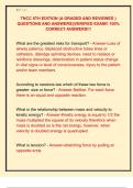
2. Hemostasis - to stop the bleeding
Hemostasis is the process of stopping the bleeding when a blood vessel wall is
damaged. If this is not the case excessive bleeding can occur.
This occurs in three steps:
1. Vasoconstriction: smooth muscle cells contract, so less blood can flow through.
(↓NO, prostacyclin ↑endothelin).
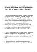
2. Primary hemostasis / Platelet plug formation: The platelets will form a plug to
temporarily seal the vessel wall --> platelet aggregation. (↓NO, prostacyclin
↑endothelin) Collagen is exposed at the site of injury that promotes platelet
adhesion Ib and IIBIIIA (receptors). the binding of platelets to collagen is stabilized
TENTAMEN PREP 1 Pagina 1
,2. Primary hemostasis / Platelet plug formation: The platelets will form a plug to
temporarily seal the vessel wall --> platelet aggregation. (↓NO, prostacyclin
↑endothelin) Collagen is exposed at the site of injury that promotes platelet
adhesion Ib and IIBIIIA (receptors). the binding of platelets to collagen is stabilized
by the Von Willebrand Factor. Platelets will release serotonin, ADP, and
thromboxane A, causing more vasoconstriction. --> Positive feedback cycle and
degranulation
3. Secondary hemostasis/ Coagulation: the formation on top of the platelet
plug --> clot formation. Fibrin is needed to strengthen the clot, forms a fibrin mesh
and this is obtained via the coagulation cascade.
Coagulation cascade can occur via the intrinsic and extrinsic pathway.
- Subendothelial collagen activates the intrinsic pathway
- The tissue factor will activate the extrinsic pathway.
Platelet factor 3 is needed for both pathways to work and a negatively charged
membrane. Coagulation enzymes are serine proteases.
Intrinsic pathway: factors for clotting are present in the blood and is triggered by
negatively charged surfaces (collagen, platelets)
Negative feedback goes via Factor V, VIII and XI
Defect in Factor VIII/IX is called hemophilia
Extrinsic pathway: requires the presence of Tissue Factor (Factor III), this is not in
the blood but is found as blood is exposed to damaged tissue, this is faster than
the intrinsic pathway.
Negative feedback goes via Factor V and VIII
Then the intrinsic and extrinsic pathways meet in the common pathway with
activated factor X. Then prothrombin can activate thrombin (Factor II) This is the
Thrombin birth. Thrombin activates fibrinogen into fibrin (Factor I) and the fibrin
mesh can now be formed. Ca++-dependent Factor XIII can also be activated,
which will form crosslinks in the now stable clot.
3. Inflammation - Neutrophils and Macrophages
Inflammation = events designed to clear cellular debris or pathogens and initiate
repair. (part of innate immunity)
Infection is the invasion of a pathogen or bacteria (inflammation ≠ infection)
Mast cells will start to release
- Histamines
- Leukotrienes
Factor XII will initiate inflammation by the activation of bradykinin.
Signs of inflammation:
1. Swelling: due to vascular permeability into the interstitial space (Cells will
contract)
2. Pain: pressure receptors in the interstitial space are triggered, which will cause
pain, bradykinin will cause pain chemically
3. Heat: due to vasodilation more blood is flowing through and blood is body
temperature therefor heat is felt.
4. Redness: due to vasodilation (histamine and leukotrienes), more blood, red
colour.
The leukocytes and Macrophages will now migrate to the site of injury. The
monocyte (macrophage) will secrete IL-1, TNF-α, IL-8 to attracts other WBCs.
Leukocytes will make contact with the vessel wall as there is slower blood flow
this is weak adhesion, but eventually will turn to firm adhesion.
(chemoattraction)
Selectins = mediate rolling of neutrophils and weak tethering
Integrins = mediate firm adhesion
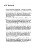
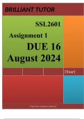
Steps of Leukocyte adhesion:
1. Rolling: in response to IL-1, TNF-α E-Selectin is expressed by adhesion
molecules. Thrombin (Factor II) will translocate P-selectin to the surface of the
endothelium. Then the leukocytes can bind to these Selectins. They bind but are
pushed by the blood flow --> and therefore they start rolling.
2. Firm adhesion & Integrin activation: leukocytes will also produce integrins
(LFA-1 and VLA-4) which are at first present in a low affinity state. Chemokines
which can bind to the chemokine receptor will change the affinity for integrins -->
integrin activation. IL-1, TNF-α will express ligands to which the LFA-1 and the
VLA-4 can bind. This will stop the rolling and the integrins change the cytoskeleton
of the leukocytes causing affinity maturation.
3. Leukocyte migration: squeeze through the junctions between the endothelial
cells. This is guided by Ca3 and Ca5 (complement factors). Diapedesis occurs
TENTAMEN PREP 1 Pagina 2
,cells. This is guided by Ca3 and Ca5 (complement factors). Diapedesis occurs
thanks to CD31/ PCAM from the blood vessel to the tissue and can migrate to the
site of injury.
Chemokines and cytokines
Chemokines = activate neutrophils and stimulate their migration.
Cytokines = activate endothelial cells to express leukocyte adhesion molecules
4. Inhibition/ Resolution of the clotting process
The first thing within the first hour is clot retraction as platelets will squeeze out
plasma to stabilize the clot. Platelet-derived growth factor (PDGF) will stimulate
the rebuild of smooth muscle and vessel walls and vascular endothelial growth
factor (VEGF) will repair the endothelial lining.
Deficiency:
- Von Willebrand Factor disease - VWF deficiency
- Bernard-Soulier syndrome - defect in GPIba
- Macrothrombocytopenia - deficiency in cytoskeleton
Fibrinolysis = the clean-up of clots that are not needed anymore this is important
as it also prevents blocking --> done by Plasmin. Plasmin is the activated form of
plasminogen and is activated by Factor XII, thrombin or protein C.
Anticoagulants = factors which inhibit clotting.
- Antithrombin, inhibits thrombin product and Factor X activation
- Protein C - inhibits Factor VIII and V
- Tissue factor pathway inhibitor (TFPI) limits tissue factor (EP)
- Plasmin, inhibits clot formation
- Prostacyclin, inhibits platelet activated
- Cl inhibitor inhibits the IP
- Alpha 1 inhibitor inhibits smooth muscle contraction.
Antiplatelet drugs:
- Clopidogrel, prasugrel, ticagrelor: target ADP
- Aspirin: targets COX
- Vorapaxar: targets thrombin receptor
- Respro, intergrin and tirofiban: block integrin AIIbB3 (aggregation)
Deficiencies can lead to bleeding or thrombosis
Keywords:
- Primary Hemostasis (platelets)
- Secondary Hemostasis (coagulation)
- Extrinsic and Intrinsic Coagulation cascades (Tissue Factor, Damaged collagen,
cleaving of coagulation factors, Factor X, Factor II (thrombin), fibrin formation and
stabilization)
- Fibrinolysis (plasmin)
- Inhibition of coagulation
- Leukocytes: myeloid (monocytes, macrophages, dendritic cells, granulocytes) and
lymphoid (T- and B-cells, NK cells)
- Inflammation (≠ infection)
- Leukocyte transmigration (selectins - rolling, chemokines - affinity maturation,
integrins – firm adhesion, junctional molecules – diapedesis)
TENTAMEN PREP 1 Pagina 3
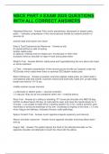
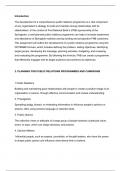
, 1. Difference arteriole & venous thrombosis
Thrombosis = a medical event that occurs when the blood clots in a wrong way block the blood flow.
1: CONSOLIDATION
Arterial thrombosis
ASSIGNMENT Venous thrombosis
zondag 15 oktober 2023 12:13
Trigger Rapture of an atherosclerotic plaque (atherosclerosis= Virchow's Triad
slagaderverkalking) 1. Hypercoagulability = increased coagulation
2. Stasis = change in blood flow (long flight on plane)
3. Endothelial injury = by drug use or trauma
Mechanism of Platelet plug formation: The platelets will form a plug to temporarily Cause:
formation seal the vessel wall --> platelet aggregation. (↓NO, prostacyclin - Low flow could lead to the accumulation of thrombin.
↑endothelin) Collagen is exposed at the site of injury that promotes - Endothelial activation of TF (clot formation)
platelet adhesion Ib and IIBIIIA (receptors). the binding of platelets to - Hypoxia (low oxygen) TF induces clot formation
collagen is stabilized by the Von Willebrand Factor. - Venous valve pockets dangerous
Then the same mechanism takes place as in Arteries --> clot
Extrinsic pathway: requires the presence of Tissue Factor (Factor III), formation
this is not in the blood but is found as blood is exposed to damaged
tissue, this is faster than the intrinsic pathway.
Then the intrinsic and extrinsic pathways meet in the common
pathway with activated factor X. Then prothrombin can activate
thrombin (Factor II) Thrombin activates fibrinogen into fibrin (Factor
I) and the fibrin mesh can now be formed. Ca++-dependent Factor XIII
can also be activated, which will form crosslinks in the now stable clot
Thrombin activates PAR1 --> more platelets recruited.
Thrombus PLATELETS (white colour) FIBRIN & RED BLOOD CELLS
composition TF --> Factor X --> thrombin --> fibrin --> trap RBC
Direction & Found in blood away from the heart Found in blood towards the heart
location Common: coronary and cerebral arteries Common: large vein of the leg , pulmonary arteries
High shear
Risk factors High blood pressure, high cholesterol, smoking, unhealthy diet and Inherited, obesity, pregnancy and immobility
age
Treatment Short term therapy, antiplatelet drugs Anticoagulant drugs, long term therapy
Could lead to: Heart attack, stroke, reduced blood flow DVT = deep vein thrombosis, blockage of pulmonary arteries
2. Treatment & prevention of arterial and venous thrombosis
Heparin is an indirect thrombin inhibitor as it stimulates prothrombin and therefor inactivates thrombin.
It is used to prevent venous thrombosis, not arterial thrombosis (rapid coagulant)
Vitamin K antagonists e.g. Warfarin block the vitamin K which makes several clotting factors, this lowers the risk
of thrombosis. This is used against venous thrombosis. (long term coagulant)
Platelet antagonist e.g. Aspirin is an antiplatelet medication and inhibits platelet aggregation (COX-1 and TXA2)
This is used in arterial thrombosis.
Fibrinolysis activators e.g. recombinant tPA and uPA activate the enzyme for plasminogen this is converted to plasmin
TENTAMEN PREP 1 Pagina 4
zondag 15 oktober 2023 11:11
1. Types of blood cells, structure, function, and origin
It starts with a hematopoietic stem cell. This then splits into the lymphoid group
and the myeloid group.
The Myeloid group
1. Red blood cells/ Enterocytes: lack a nucleus and organelles and contain
haemoglobin (oxygen transport and gas exchange). The production of RBCs
Erythropoiesis is driven by low oxygen levels in the tissues and occurs in the red
bone marrow (where the hematopoietic stem cell is present)
2. Mast cells: mediate host defence against pathogens by releasing granules and
are active in allergic reactions
3. White blood cells/ Leukocytes: contain a nucleus and organelles. The overall
function is protection against diseases. Leukopoiesis is stimulated by interleukins.
They can be subdivided into
3.1 Granulocytes: bigger than RBCs, but live shorter, are all phagocytes to a
certain degree. Contain:
- Neutrophils: responsible for defence against bacterial infections by
phagocytosis. (most present WBC)
○ Band neutrophils = young neutrophils, 1 nucleus particle
○ Segmented neutrophils = older neutrophil, multiple nucleus particles.
- Eosinophils: responsible for fighting allergic reactions and parasitic
infections. They can be recognized by the red colour and bi-lobed nucleus
- Basophils: are filled with Histamine, which acts as a vasodilator and attracts
other WBC to the inflamed site.
3.2 Agranulocytes: lack cytoplasmatic granules their nuclei are kidney shaped.
- Monocytes are the largest cell in the blood , they can differentiate into
macrophages and protect from viruses, bacteria and chronic infections. And
are important in activating lymphocytes.
- Lymphocytes (lymphoid group): play a crucial role in immunity by direct cell
attach or via antibodies
○ B-lymphocytes: give rise to plasma cells which will produce
antibodies.
○ T-lymphocytes: act directly against virus or tumour cells
○ Natural killer cells: help in the innate immune system
4. Platelets/ Thrombocytes: are fragments of Megakaryocytes which are filled
with granules needed for the clotting process to form the temporary plug. The
formation of platelets is determined by thrombopoietin.
Thrombopoiesis:
1. MK develops in the bone marrow
2. endomitosis to create a polyploid nucleus (MK are polyploids)
3. cytoplasmic maturation
4. proplatelet formation and release
5. pre platelet to proplatelet interconversion
6. platelet release
Platelets are stimulated and then Ca++ is released, this amplifies its own reaction
and this is how platelets signal
Platelet adhesion is dependent on flow. In the middle of the vessel the flow is
higher than on the sides. Shear is the difference in flow between the layers. Shear
is minimal in the middle had highest near the edges.
2. Hemostasis - to stop the bleeding
Hemostasis is the process of stopping the bleeding when a blood vessel wall is
damaged. If this is not the case excessive bleeding can occur.
This occurs in three steps:
1. Vasoconstriction: smooth muscle cells contract, so less blood can flow through.
(↓NO, prostacyclin ↑endothelin).
2. Primary hemostasis / Platelet plug formation: The platelets will form a plug to
temporarily seal the vessel wall --> platelet aggregation. (↓NO, prostacyclin
↑endothelin) Collagen is exposed at the site of injury that promotes platelet
adhesion Ib and IIBIIIA (receptors). the binding of platelets to collagen is stabilized
TENTAMEN PREP 1 Pagina 1
,2. Primary hemostasis / Platelet plug formation: The platelets will form a plug to
temporarily seal the vessel wall --> platelet aggregation. (↓NO, prostacyclin
↑endothelin) Collagen is exposed at the site of injury that promotes platelet
adhesion Ib and IIBIIIA (receptors). the binding of platelets to collagen is stabilized
by the Von Willebrand Factor. Platelets will release serotonin, ADP, and
thromboxane A, causing more vasoconstriction. --> Positive feedback cycle and
degranulation
3. Secondary hemostasis/ Coagulation: the formation on top of the platelet
plug --> clot formation. Fibrin is needed to strengthen the clot, forms a fibrin mesh
and this is obtained via the coagulation cascade.
Coagulation cascade can occur via the intrinsic and extrinsic pathway.
- Subendothelial collagen activates the intrinsic pathway
- The tissue factor will activate the extrinsic pathway.
Platelet factor 3 is needed for both pathways to work and a negatively charged
membrane. Coagulation enzymes are serine proteases.
Intrinsic pathway: factors for clotting are present in the blood and is triggered by
negatively charged surfaces (collagen, platelets)
Negative feedback goes via Factor V, VIII and XI
Defect in Factor VIII/IX is called hemophilia
Extrinsic pathway: requires the presence of Tissue Factor (Factor III), this is not in
the blood but is found as blood is exposed to damaged tissue, this is faster than
the intrinsic pathway.
Negative feedback goes via Factor V and VIII
Then the intrinsic and extrinsic pathways meet in the common pathway with
activated factor X. Then prothrombin can activate thrombin (Factor II) This is the
Thrombin birth. Thrombin activates fibrinogen into fibrin (Factor I) and the fibrin
mesh can now be formed. Ca++-dependent Factor XIII can also be activated,
which will form crosslinks in the now stable clot.
3. Inflammation - Neutrophils and Macrophages
Inflammation = events designed to clear cellular debris or pathogens and initiate
repair. (part of innate immunity)
Infection is the invasion of a pathogen or bacteria (inflammation ≠ infection)
Mast cells will start to release
- Histamines
- Leukotrienes
Factor XII will initiate inflammation by the activation of bradykinin.
Signs of inflammation:
1. Swelling: due to vascular permeability into the interstitial space (Cells will
contract)
2. Pain: pressure receptors in the interstitial space are triggered, which will cause
pain, bradykinin will cause pain chemically
3. Heat: due to vasodilation more blood is flowing through and blood is body
temperature therefor heat is felt.
4. Redness: due to vasodilation (histamine and leukotrienes), more blood, red
colour.
The leukocytes and Macrophages will now migrate to the site of injury. The
monocyte (macrophage) will secrete IL-1, TNF-α, IL-8 to attracts other WBCs.
Leukocytes will make contact with the vessel wall as there is slower blood flow
this is weak adhesion, but eventually will turn to firm adhesion.
(chemoattraction)
Selectins = mediate rolling of neutrophils and weak tethering
Integrins = mediate firm adhesion
Steps of Leukocyte adhesion:
1. Rolling: in response to IL-1, TNF-α E-Selectin is expressed by adhesion
molecules. Thrombin (Factor II) will translocate P-selectin to the surface of the
endothelium. Then the leukocytes can bind to these Selectins. They bind but are
pushed by the blood flow --> and therefore they start rolling.
2. Firm adhesion & Integrin activation: leukocytes will also produce integrins
(LFA-1 and VLA-4) which are at first present in a low affinity state. Chemokines
which can bind to the chemokine receptor will change the affinity for integrins -->
integrin activation. IL-1, TNF-α will express ligands to which the LFA-1 and the
VLA-4 can bind. This will stop the rolling and the integrins change the cytoskeleton
of the leukocytes causing affinity maturation.
3. Leukocyte migration: squeeze through the junctions between the endothelial
cells. This is guided by Ca3 and Ca5 (complement factors). Diapedesis occurs
TENTAMEN PREP 1 Pagina 2
,cells. This is guided by Ca3 and Ca5 (complement factors). Diapedesis occurs
thanks to CD31/ PCAM from the blood vessel to the tissue and can migrate to the
site of injury.
Chemokines and cytokines
Chemokines = activate neutrophils and stimulate their migration.
Cytokines = activate endothelial cells to express leukocyte adhesion molecules
4. Inhibition/ Resolution of the clotting process
The first thing within the first hour is clot retraction as platelets will squeeze out
plasma to stabilize the clot. Platelet-derived growth factor (PDGF) will stimulate
the rebuild of smooth muscle and vessel walls and vascular endothelial growth
factor (VEGF) will repair the endothelial lining.
Deficiency:
- Von Willebrand Factor disease - VWF deficiency
- Bernard-Soulier syndrome - defect in GPIba
- Macrothrombocytopenia - deficiency in cytoskeleton
Fibrinolysis = the clean-up of clots that are not needed anymore this is important
as it also prevents blocking --> done by Plasmin. Plasmin is the activated form of
plasminogen and is activated by Factor XII, thrombin or protein C.
Anticoagulants = factors which inhibit clotting.
- Antithrombin, inhibits thrombin product and Factor X activation
- Protein C - inhibits Factor VIII and V
- Tissue factor pathway inhibitor (TFPI) limits tissue factor (EP)
- Plasmin, inhibits clot formation
- Prostacyclin, inhibits platelet activated
- Cl inhibitor inhibits the IP
- Alpha 1 inhibitor inhibits smooth muscle contraction.
Antiplatelet drugs:
- Clopidogrel, prasugrel, ticagrelor: target ADP
- Aspirin: targets COX
- Vorapaxar: targets thrombin receptor
- Respro, intergrin and tirofiban: block integrin AIIbB3 (aggregation)
Deficiencies can lead to bleeding or thrombosis
Keywords:
- Primary Hemostasis (platelets)
- Secondary Hemostasis (coagulation)
- Extrinsic and Intrinsic Coagulation cascades (Tissue Factor, Damaged collagen,
cleaving of coagulation factors, Factor X, Factor II (thrombin), fibrin formation and
stabilization)
- Fibrinolysis (plasmin)
- Inhibition of coagulation
- Leukocytes: myeloid (monocytes, macrophages, dendritic cells, granulocytes) and
lymphoid (T- and B-cells, NK cells)
- Inflammation (≠ infection)
- Leukocyte transmigration (selectins - rolling, chemokines - affinity maturation,
integrins – firm adhesion, junctional molecules – diapedesis)
TENTAMEN PREP 1 Pagina 3
, 1. Difference arteriole & venous thrombosis
Thrombosis = a medical event that occurs when the blood clots in a wrong way block the blood flow.
1: CONSOLIDATION
Arterial thrombosis
ASSIGNMENT Venous thrombosis
zondag 15 oktober 2023 12:13
Trigger Rapture of an atherosclerotic plaque (atherosclerosis= Virchow's Triad
slagaderverkalking) 1. Hypercoagulability = increased coagulation
2. Stasis = change in blood flow (long flight on plane)
3. Endothelial injury = by drug use or trauma
Mechanism of Platelet plug formation: The platelets will form a plug to temporarily Cause:
formation seal the vessel wall --> platelet aggregation. (↓NO, prostacyclin - Low flow could lead to the accumulation of thrombin.
↑endothelin) Collagen is exposed at the site of injury that promotes - Endothelial activation of TF (clot formation)
platelet adhesion Ib and IIBIIIA (receptors). the binding of platelets to - Hypoxia (low oxygen) TF induces clot formation
collagen is stabilized by the Von Willebrand Factor. - Venous valve pockets dangerous
Then the same mechanism takes place as in Arteries --> clot
Extrinsic pathway: requires the presence of Tissue Factor (Factor III), formation
this is not in the blood but is found as blood is exposed to damaged
tissue, this is faster than the intrinsic pathway.
Then the intrinsic and extrinsic pathways meet in the common
pathway with activated factor X. Then prothrombin can activate
thrombin (Factor II) Thrombin activates fibrinogen into fibrin (Factor
I) and the fibrin mesh can now be formed. Ca++-dependent Factor XIII
can also be activated, which will form crosslinks in the now stable clot
Thrombin activates PAR1 --> more platelets recruited.
Thrombus PLATELETS (white colour) FIBRIN & RED BLOOD CELLS
composition TF --> Factor X --> thrombin --> fibrin --> trap RBC
Direction & Found in blood away from the heart Found in blood towards the heart
location Common: coronary and cerebral arteries Common: large vein of the leg , pulmonary arteries
High shear
Risk factors High blood pressure, high cholesterol, smoking, unhealthy diet and Inherited, obesity, pregnancy and immobility
age
Treatment Short term therapy, antiplatelet drugs Anticoagulant drugs, long term therapy
Could lead to: Heart attack, stroke, reduced blood flow DVT = deep vein thrombosis, blockage of pulmonary arteries
2. Treatment & prevention of arterial and venous thrombosis
Heparin is an indirect thrombin inhibitor as it stimulates prothrombin and therefor inactivates thrombin.
It is used to prevent venous thrombosis, not arterial thrombosis (rapid coagulant)
Vitamin K antagonists e.g. Warfarin block the vitamin K which makes several clotting factors, this lowers the risk
of thrombosis. This is used against venous thrombosis. (long term coagulant)
Platelet antagonist e.g. Aspirin is an antiplatelet medication and inhibits platelet aggregation (COX-1 and TXA2)
This is used in arterial thrombosis.
Fibrinolysis activators e.g. recombinant tPA and uPA activate the enzyme for plasminogen this is converted to plasmin
TENTAMEN PREP 1 Pagina 4