Tendons
Introduction
Highly aligned tensile tissue attaching muscle to bone
Allow muscles to act at a distance from a joint Abreviations:
In general: act to transmit tensile forces between muscle and bone Mfs= muscle fibres
Store elastic energy
Dominated by ECM
mechanosensitive
Importance of tendons:
Structure and functions- Benjamin Tendon can rupture
Intermediate tendon- connects one muscle belly to another spontaneously
Intramuscular tendons- allows MFs to have a penate arrangement
collagen network of the perimysium that forms the basis for the Prone to pathology –
mechanical link between tendon and muscle fibres and this is promoted especially in athletes,
by specialized ‘perimysial junctional plates both elite and
tendon ECM= largely that of a dense fibrous connective tissue recreational. Tendon
Aponeuroses= flattened tendons injury affects 6% of
the increasing importance of tissue engineering and stem cell biology in population & up to
biomedical science has raised interest in creating artificial tendons or in 50% of athletes
using mesenchymal stem cells to promote repair (Zhang & Chang, 2003; Tendinopathy: chronic
Smith & Webbon, 2005 injury due to tendon
Tendon Structure overuse injury
The largest tendon is the Achilles and its shape varies from proximal to Partial or complete
distal as it approaches its calcaneal attachment site tendon ruptures lead to
Usually, extensor tendons are more flattened than flexor tendons – which loss or reuction in
tend to be round or oval mobility
tendons largely consist of collagens and proteoglycans and are dominated
by the fibril-forming, type I collagen. However, other collagens (e.g. II,
III, V, VI, IX, XI) are also present (Fukuta et al. 1998; Ottani et al. 2002;
Kjaer, 2004). Tendon disorders are:
Proteoglycans are primarily responsible for the viscoelastic behaviour of Very common.
tendons, but do not make any major contribution to their tensile strength ( Tendon injuries
The principal role of the collagen fibres is to resist tension, although they represent approximately
still allow for a certain degree of compliance (i.e. reversible longitudinal 50% of all sports
deformation) injuries
One of the important features in tendons is the ability of their fascicles to Have an increasing
slide independently against each other. This allows them to transmit prevalence with age
tension despite the changing angles of a joint as it moves (Fallon et al. Have a substantial effect
2002) and allows tendons to change shape as their muscles contract. on quality of life
Tendon Cells Considerable economic
Tenocyte- longitudinal rows, near the collagen fibrils burden on health care
Aging- cells flatten and become less numerous and their long, thin systems
cytoplasmic projections shorten and diminish in number
Mature tendon cells thus have a complex system of sheet-like and finger-
like processes that facilitate intercellular communication via gap Development of effective
junctions in a way that is comparable to the communication between therapeutics is hindered by the
osteocytes in bone lack of fundamental data on
Marker: transcription factor scleraxis has been used to identify tendon or the biology of:
ligament cells at all stages of their development Tendon development
Marker: tenomodulin – a molecule whose expression is induced by Signal transduction
scleraxis Mechanotransduction
Marker: tenascin-C. This is expressed by tenocytes in response to and basic mechanisms
mechanical stress, but again is not specific for tendons alone, for it is also underlying tendon
present in bone, smooth muscle and healing fibroblasts pathogenesis and
healing
, • Tendons considered to have a
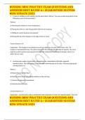
Tendon Structure poor blood supply
Tendon transmits muscle contraction force to bone • Paratenon-covered tendons:
To maintain posture or produce motion vessels from surrounding tissue
Important for you to consider how the structure and composition penetrate at any point along tendon
of tendon permits such force transmission (sheathed tendons avascular by
comparison)
• No nerve fibres within tendon
body
• Paratenon and epitenon contains
nerve endings (sensory – pain
receptors)
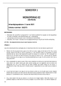
• Hierarchical arrangement
• Fibrils: Principle tensile element
in tendon.
• Built in safety mechanism against
Hierarchical arrangement failure
Triple helix type 1 collagen molecules (tropocollagen) • Offers the possibility that local
Assembled in fibrils fibres fascicles tendon unit failure can be isolated within one
Not all tendons have a paratenon, your energy storing tendons do part of the cross-sectional profile of
a tendon and not spread throughout
its thickness
• The ability of fibrils, fibres and/or
fascicles to move relative to each
other is essential in allowing them
to lengthen/change shape.
• Type 1 collagen and other ECM
molecules are produced by tendon
cells: Tenocytes=tendinocytes
=tendon fibroblasts
• These cells are interspersed
between collagen fibres and within
the endotenon
• Cytoplasmic processes are used to
communicate with other cells and
Type I collagen- most important respond to loading
Very little is known about the mechanisms driving the spatial
organization.
tenocytes: Tendon function
- Need to express of Col1a1 and Col1a2 to be able to profuce • All tendons perform a positional
collagen role – loaded along their long axis in
- Need to express the transcription factors such as Scleraxis tension.
- Scleraxis regulates the expression of Tenomodulin • Enables the muscles to move the
Tenomodulin is a marker for mature tenocytes skeleton
Tenocytes also need to secrete growth factors which are known to • Some tendons have an additional
promote collagen expression and synthesis in tendon tissue during role; stretching when loaded to store
development and in adult life; TGF-β and FGF’s Mechanical energy ‘Energy storing tendons’
forces are also involved in type 1 collagen synthesis • Improves the efficiency of
locomotion
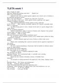
• Energy storing tendon is more
• Compositional differences between energy storing tendons and prone to injury!
positional tendons • Energy storing tendons are less
• Higher GAG/PG content in energy storing tendons stiff and more extensible that
• Energy storing tendons rely on lubricin and elastin between fascicles positional tendons i.e. more ‘elastic’
to enable a more elastic, recoverable fascicle sliding • Springs in locomotion
• Energy storing tendons also appear to be helically arranged • Absorb and release force in a
contributing to the spring like elastic behaviour spring like manner
Introduction
Highly aligned tensile tissue attaching muscle to bone
Allow muscles to act at a distance from a joint Abreviations:
In general: act to transmit tensile forces between muscle and bone Mfs= muscle fibres
Store elastic energy
Dominated by ECM
mechanosensitive
Importance of tendons:
Structure and functions- Benjamin Tendon can rupture
Intermediate tendon- connects one muscle belly to another spontaneously
Intramuscular tendons- allows MFs to have a penate arrangement
collagen network of the perimysium that forms the basis for the Prone to pathology –
mechanical link between tendon and muscle fibres and this is promoted especially in athletes,
by specialized ‘perimysial junctional plates both elite and
tendon ECM= largely that of a dense fibrous connective tissue recreational. Tendon
Aponeuroses= flattened tendons injury affects 6% of
the increasing importance of tissue engineering and stem cell biology in population & up to
biomedical science has raised interest in creating artificial tendons or in 50% of athletes
using mesenchymal stem cells to promote repair (Zhang & Chang, 2003; Tendinopathy: chronic
Smith & Webbon, 2005 injury due to tendon
Tendon Structure overuse injury
The largest tendon is the Achilles and its shape varies from proximal to Partial or complete
distal as it approaches its calcaneal attachment site tendon ruptures lead to
Usually, extensor tendons are more flattened than flexor tendons – which loss or reuction in
tend to be round or oval mobility
tendons largely consist of collagens and proteoglycans and are dominated
by the fibril-forming, type I collagen. However, other collagens (e.g. II,
III, V, VI, IX, XI) are also present (Fukuta et al. 1998; Ottani et al. 2002;
Kjaer, 2004). Tendon disorders are:
Proteoglycans are primarily responsible for the viscoelastic behaviour of Very common.
tendons, but do not make any major contribution to their tensile strength ( Tendon injuries
The principal role of the collagen fibres is to resist tension, although they represent approximately
still allow for a certain degree of compliance (i.e. reversible longitudinal 50% of all sports
deformation) injuries
One of the important features in tendons is the ability of their fascicles to Have an increasing
slide independently against each other. This allows them to transmit prevalence with age
tension despite the changing angles of a joint as it moves (Fallon et al. Have a substantial effect
2002) and allows tendons to change shape as their muscles contract. on quality of life
Tendon Cells Considerable economic
Tenocyte- longitudinal rows, near the collagen fibrils burden on health care
Aging- cells flatten and become less numerous and their long, thin systems
cytoplasmic projections shorten and diminish in number
Mature tendon cells thus have a complex system of sheet-like and finger-
like processes that facilitate intercellular communication via gap Development of effective
junctions in a way that is comparable to the communication between therapeutics is hindered by the
osteocytes in bone lack of fundamental data on
Marker: transcription factor scleraxis has been used to identify tendon or the biology of:
ligament cells at all stages of their development Tendon development
Marker: tenomodulin – a molecule whose expression is induced by Signal transduction
scleraxis Mechanotransduction
Marker: tenascin-C. This is expressed by tenocytes in response to and basic mechanisms
mechanical stress, but again is not specific for tendons alone, for it is also underlying tendon
present in bone, smooth muscle and healing fibroblasts pathogenesis and
healing
, • Tendons considered to have a
Tendon Structure poor blood supply
Tendon transmits muscle contraction force to bone • Paratenon-covered tendons:
To maintain posture or produce motion vessels from surrounding tissue
Important for you to consider how the structure and composition penetrate at any point along tendon
of tendon permits such force transmission (sheathed tendons avascular by
comparison)
• No nerve fibres within tendon
body
• Paratenon and epitenon contains
nerve endings (sensory – pain
receptors)
• Hierarchical arrangement
• Fibrils: Principle tensile element
in tendon.
• Built in safety mechanism against
Hierarchical arrangement failure
Triple helix type 1 collagen molecules (tropocollagen) • Offers the possibility that local
Assembled in fibrils fibres fascicles tendon unit failure can be isolated within one
Not all tendons have a paratenon, your energy storing tendons do part of the cross-sectional profile of
a tendon and not spread throughout
its thickness
• The ability of fibrils, fibres and/or
fascicles to move relative to each
other is essential in allowing them
to lengthen/change shape.
• Type 1 collagen and other ECM
molecules are produced by tendon
cells: Tenocytes=tendinocytes
=tendon fibroblasts
• These cells are interspersed
between collagen fibres and within
the endotenon
• Cytoplasmic processes are used to
communicate with other cells and
Type I collagen- most important respond to loading
Very little is known about the mechanisms driving the spatial
organization.
tenocytes: Tendon function
- Need to express of Col1a1 and Col1a2 to be able to profuce • All tendons perform a positional
collagen role – loaded along their long axis in
- Need to express the transcription factors such as Scleraxis tension.
- Scleraxis regulates the expression of Tenomodulin • Enables the muscles to move the
Tenomodulin is a marker for mature tenocytes skeleton
Tenocytes also need to secrete growth factors which are known to • Some tendons have an additional
promote collagen expression and synthesis in tendon tissue during role; stretching when loaded to store
development and in adult life; TGF-β and FGF’s Mechanical energy ‘Energy storing tendons’
forces are also involved in type 1 collagen synthesis • Improves the efficiency of
locomotion
• Energy storing tendon is more
• Compositional differences between energy storing tendons and prone to injury!
positional tendons • Energy storing tendons are less
• Higher GAG/PG content in energy storing tendons stiff and more extensible that
• Energy storing tendons rely on lubricin and elastin between fascicles positional tendons i.e. more ‘elastic’
to enable a more elastic, recoverable fascicle sliding • Springs in locomotion
• Energy storing tendons also appear to be helically arranged • Absorb and release force in a
contributing to the spring like elastic behaviour spring like manner