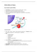
Protein folding and disease
Key concepts in protein folding
• Central dogma – flow of genetic information from RNA to DNA ® proteins
• Transcription (RNA synthesis) and translation (protein synthesis)
• mRNA leaves the nucleus to be translated into proteins in the cytosol
• Growing polypeptide chain is formed – peptide chain is transferred from resident tRNA to
incoming tRNA
Life cycle of a protein
• DNA ® RNA ® protein ® folding ® (post-translational modification/s) ® Transport to specific
or extracellular compartments ® Functional protein ® Degradation
• PTM –
o Proteolytic cleavage
o Disulfide bonding
o Phosphorylation
o Acetylation
o N and O-linked glycosylation
o Lipidation
• Amino acid sequence is an immature stage of the protein
o Needs to be developed to perform with the correct three-dimensional conformation at
the correct location
o Movement from the cytosol to its correct spot
• Protein folding – undergone to reach the desired structure ® native protein
• Degradation – proteolysis of damaged proteins or proteins at the end of life-span
,Where do newly synthesised proteins fold?
• The rough endoplasmic reticulum, begins folding in the cytoplasm but can also fold in the
cytoplasm
o Every single newly synthesised protein begins in the cytoplasm before it is shuttled into
the ER
o When it starts being made in cytoplasm ® folding begins
Why is protein folding important?
• Protein structure is critical to function – different proteins have similar structures but perform
different functions due to folding
• Helps protein to reach its biologically active 3D conformation
• Stabilises the protein by minimising free energy
o Protein folds in such a way that it is spontaneous
o Thermodynamically favourable process by minimising free energy
• Regulates activity and signalling
o Creates binding sites, allowing for the binding of other proteins or ligands
What is the protein folding process?
Protein structures
• Primary protein structure – sequences of a chain of amino acids, joined
by peptide bonds
• Secondary protein structure – hydrogen bonding of the peptide
backbone causes the amino acids to fold into a repeating pattern
• Tertiary protein structure – three-
dimensional folding pattern of a protein Hydrophobic interactions
due to side chain interactions Disulphide bridges
• Quaternary protein structure – protein Wan der Waal forces
consisting of ≥ amino acids chain
How does a protein know in what 3D conformation to fold?
• Anfinsen experiment – determination that the information required for
protein folding is contained with primary sequence of the amino acid
sequence
• Treat ribonuclease with either urea (disrupts H bonds) or beta-mercaptoethanol (disrupts
disulphide bridges)
• Results in complete degradation of RNase A ® returns to primary structure ® inactive
, • Removal of these solutions allows RNase to refold, spontaneous
o (1) When treated with urea but not ME = created inactive/ scrambled RNase
A with randomly formed disulfide bonds
o Absence of hydrogen bonds
o (2) When trace amounts of ME are added ® disulphide bonds interact at
correct positions (native ribonuclease A)
o Randomly formed disulfide bonds break up, allowing normal bonds to form
o By the absence of urea, able to form correct disulphide bonding
• Conclusions
o Information for forming the mature 3D structure of a protein is determined by its primary
structure
o Folding proceeds in ordered states – information in primary sequence drives H-bonding
in secondary ® drives correct formation of disulphide bonds in 3D state
o Proteins fold to reach the thermodynamically most stable state
How do newly translated proteins fold in vivo?
• Hydrophobic effect – In aqueous solutions, proteins fold the hydrophobic areas away from the
hydrophilic solution
• Protein folding occurs in a region of high [protein] (many proteins folding at the same time) ®
form aggregates with each through hydrophobic interactions
o Continue to grow
• To prevent this problem, protein folding is assisted by chaperones/ chaperonins
• Note: do not provide any additional information for folding
o Prevents non-specific hydrophobic interactions
o Provides a chamber in which proteins can fold in isolation (chaperonins)
o Stabilises unfolded proteins during transport to subcellular organelle
• Primary sequence interacts with chaperone and then chaperonin before it reaches its native state
• As protein synthesis continues, more chaperones come and bind to hydrophobic areas, allowing
protein to bind to three-dimensional conformation (co-translational)
, • Chaperonin provides an isolated chamber in which the protein can fold if chaperone-assisted
binding is not sufficient
o Thus it needs to be fully translated by the time it reaches the chaperonin
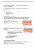
HSP70 in eukaryotes
• Hsp70 has three distinct domains = ATPase domain, substrate binding domain, Lid region
o Linker region between ATPase domain and SBD
• ATP-driven process
• Substrate binding domain – binds to the hydrophobic region of the polypeptide chain
• Lid region – closes over the hydrophobic pocket, holding the chain in place
o Allows the chaperone to stay in contact with the polypeptide chain
• Function is an open and closed format
o No contact with protein = open conformation
• Chaperone makes contact with the hydrophobic cleft ® conformational change ® Lid region
clamps the protein
• While different chaperones are bound, the protein is allowed to fold
• Closed conformation by the hydrolysis of ATP
HSP60 – a chaperonin
• Hollow chamber whereby protein folding can occur in isolation
• GroEL = barrel of the chaperonin
o Contains hydrophobic binding sites
• GroES = lid
• Functions in an open and closed format
• Nascent polypeptide chain makes contact ® causes GroES to clamp down on GroEL
• Provides energy for the chain to unfold or push it further into the barrow to allow it to fold in
isolation
• Unfolded/ partially folded due to improperly folded state by chaperone
• ATP – (1) provides energy for the unfolding of the protein, giving it a chance to refold again (2)
facilitates the binding of GroEL
What happens if protein folding mechanisms fail?
• Proteins that fail to fold must be delivered
to the proteasome for proteolysis
o Misfolding of protein = not active
or not correct conformation
• Proteasome – two cap regions on their side
of the core region
o Core – catalytic unit, location of
proteolysis
o Cap – protein recognition +
unfolding mechanism + feeding the protein into the core unit
• Proteins are tagged for degradation in the proteasome with ubiquitin
Key concepts in protein folding
• Central dogma – flow of genetic information from RNA to DNA ® proteins
• Transcription (RNA synthesis) and translation (protein synthesis)
• mRNA leaves the nucleus to be translated into proteins in the cytosol
• Growing polypeptide chain is formed – peptide chain is transferred from resident tRNA to
incoming tRNA
Life cycle of a protein
• DNA ® RNA ® protein ® folding ® (post-translational modification/s) ® Transport to specific
or extracellular compartments ® Functional protein ® Degradation
• PTM –
o Proteolytic cleavage
o Disulfide bonding
o Phosphorylation
o Acetylation
o N and O-linked glycosylation
o Lipidation
• Amino acid sequence is an immature stage of the protein
o Needs to be developed to perform with the correct three-dimensional conformation at
the correct location
o Movement from the cytosol to its correct spot
• Protein folding – undergone to reach the desired structure ® native protein
• Degradation – proteolysis of damaged proteins or proteins at the end of life-span
,Where do newly synthesised proteins fold?
• The rough endoplasmic reticulum, begins folding in the cytoplasm but can also fold in the
cytoplasm
o Every single newly synthesised protein begins in the cytoplasm before it is shuttled into
the ER
o When it starts being made in cytoplasm ® folding begins
Why is protein folding important?
• Protein structure is critical to function – different proteins have similar structures but perform
different functions due to folding
• Helps protein to reach its biologically active 3D conformation
• Stabilises the protein by minimising free energy
o Protein folds in such a way that it is spontaneous
o Thermodynamically favourable process by minimising free energy
• Regulates activity and signalling
o Creates binding sites, allowing for the binding of other proteins or ligands
What is the protein folding process?
Protein structures
• Primary protein structure – sequences of a chain of amino acids, joined
by peptide bonds
• Secondary protein structure – hydrogen bonding of the peptide
backbone causes the amino acids to fold into a repeating pattern
• Tertiary protein structure – three-
dimensional folding pattern of a protein Hydrophobic interactions
due to side chain interactions Disulphide bridges
• Quaternary protein structure – protein Wan der Waal forces
consisting of ≥ amino acids chain
How does a protein know in what 3D conformation to fold?
• Anfinsen experiment – determination that the information required for
protein folding is contained with primary sequence of the amino acid
sequence
• Treat ribonuclease with either urea (disrupts H bonds) or beta-mercaptoethanol (disrupts
disulphide bridges)
• Results in complete degradation of RNase A ® returns to primary structure ® inactive
, • Removal of these solutions allows RNase to refold, spontaneous
o (1) When treated with urea but not ME = created inactive/ scrambled RNase
A with randomly formed disulfide bonds
o Absence of hydrogen bonds
o (2) When trace amounts of ME are added ® disulphide bonds interact at
correct positions (native ribonuclease A)
o Randomly formed disulfide bonds break up, allowing normal bonds to form
o By the absence of urea, able to form correct disulphide bonding
• Conclusions
o Information for forming the mature 3D structure of a protein is determined by its primary
structure
o Folding proceeds in ordered states – information in primary sequence drives H-bonding
in secondary ® drives correct formation of disulphide bonds in 3D state
o Proteins fold to reach the thermodynamically most stable state
How do newly translated proteins fold in vivo?
• Hydrophobic effect – In aqueous solutions, proteins fold the hydrophobic areas away from the
hydrophilic solution
• Protein folding occurs in a region of high [protein] (many proteins folding at the same time) ®
form aggregates with each through hydrophobic interactions
o Continue to grow
• To prevent this problem, protein folding is assisted by chaperones/ chaperonins
• Note: do not provide any additional information for folding
o Prevents non-specific hydrophobic interactions
o Provides a chamber in which proteins can fold in isolation (chaperonins)
o Stabilises unfolded proteins during transport to subcellular organelle
• Primary sequence interacts with chaperone and then chaperonin before it reaches its native state
• As protein synthesis continues, more chaperones come and bind to hydrophobic areas, allowing
protein to bind to three-dimensional conformation (co-translational)
, • Chaperonin provides an isolated chamber in which the protein can fold if chaperone-assisted
binding is not sufficient
o Thus it needs to be fully translated by the time it reaches the chaperonin
HSP70 in eukaryotes
• Hsp70 has three distinct domains = ATPase domain, substrate binding domain, Lid region
o Linker region between ATPase domain and SBD
• ATP-driven process
• Substrate binding domain – binds to the hydrophobic region of the polypeptide chain
• Lid region – closes over the hydrophobic pocket, holding the chain in place
o Allows the chaperone to stay in contact with the polypeptide chain
• Function is an open and closed format
o No contact with protein = open conformation
• Chaperone makes contact with the hydrophobic cleft ® conformational change ® Lid region
clamps the protein
• While different chaperones are bound, the protein is allowed to fold
• Closed conformation by the hydrolysis of ATP
HSP60 – a chaperonin
• Hollow chamber whereby protein folding can occur in isolation
• GroEL = barrel of the chaperonin
o Contains hydrophobic binding sites
• GroES = lid
• Functions in an open and closed format
• Nascent polypeptide chain makes contact ® causes GroES to clamp down on GroEL
• Provides energy for the chain to unfold or push it further into the barrow to allow it to fold in
isolation
• Unfolded/ partially folded due to improperly folded state by chaperone
• ATP – (1) provides energy for the unfolding of the protein, giving it a chance to refold again (2)
facilitates the binding of GroEL
What happens if protein folding mechanisms fail?
• Proteins that fail to fold must be delivered
to the proteasome for proteolysis
o Misfolding of protein = not active
or not correct conformation
• Proteasome – two cap regions on their side
of the core region
o Core – catalytic unit, location of
proteolysis
o Cap – protein recognition +
unfolding mechanism + feeding the protein into the core unit
• Proteins are tagged for degradation in the proteasome with ubiquitin