Ne#omuscul* Junction
• Neuromuscular junction (NMJ) / myoneural junction – the place where the
motor neuron makes a functional contact with the skeletal muscle fibre
o Specialised force of a chemical synapse
o Comprised of alpha motor neuron and the muscle fibre it
innervates
• Nerves control skeletal muscle contractions
• Muscles that require a precise control have a nerve: myofibril ratio of one
to one
o E.g. ocular muscle – muscle associated with the eye
• Other muscles can have one nerve fibre that innervates many myofibrils
o E.g. latissimus muscle – occupies most of the lower posterior
thorax
Physiology
Neuromuscular junction Synapse
Exists between a motor neuron and a skeletal A junction between neurones or other tissue
muscle
A junction will always respond to an action Junctions between neurons or between neurons
potential in the presynaptic nerve and other cells
Has a safety factor – sufficient release of Enables communication with the nervous system
neurotransmitter to ensure actional potential + between neurons and target cells in various
generation in the effector organ tissues
• Redundancy helps prevent failure in muscle
activation
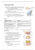
Terminal boutons
• The terminal inflated portion of the axon, containing
specialised apparatus to release neurotransmitters
o The axon terminus is the whole region of thickening,
the terminal bouton is the specialised region
• Myelin sheath surrounding the motor axons ends near the
surface of the muscle fibre
• Axon divides into a number of short processes that lie
embedded in grooves on the muscle-fibre surface
• Location of ACh storage and release
• Synaptic cleft/ cleft – space between axon terminal and
synaptic trough
o 20 to 30 nm wide
o Space filled with ECF – gel of carbohydrate-rich
amorphous material
, • Motor end plate – plasma membrane opposite the terminal bout
o Invaginated and contained nicotinic cholinergic receptors (nAChRs)
Synaptic cleft
• Folds of the synaptic trough
• Increase post-synaptic surface area
• Location of majority of Ach receptor = ligand-
gated Na+ and K+ channels
• Post-synaptic membrane contains
acetylcholinesterase – splits Ach ® acetyl-CoA
and choline
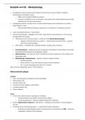
1. Arrival of action potential at the terminal bouton
2. Depolarisation causes opening of Ca2+ channels and the influx of calcium ions into the cell
3. Triggers the release of ACh from vesicles by exocytosis
• Four calcium ions trigger the release of one ACh vesicle for exocytosis
ACh receptors
4. ACh is release into the synaptic cleft
5. ACh binds to nAChRs at the motor end plate ® opening ion channels
Nicotinic ACh receptors
• 2 alpha, 1 beta, 1 delta and gamma
• Two ACh molecules attach to two alpha subunits ® opens the channel
• Resting state = two ACh molecules are not attached to the alpha subunits ® channel remains closed
End plate potentials
6. Na+ flows into the muscle cell and causes an end plate potential, sufficient to trigger an action
potential
7. The flow of Na+ generates an action potential which travels to the myofibril ® muscle contraction
Excitatory post-synaptic potentials
8. The interaction of ACh with its receptors in the post-synaptic membrane opens the chemically
regulated gates ® depolarisation of this region in the membrane
9. Depolarisation produced = excitatory postsynaptic potentials (EPSP)
• Neuromuscular junction (NMJ) / myoneural junction – the place where the
motor neuron makes a functional contact with the skeletal muscle fibre
o Specialised force of a chemical synapse
o Comprised of alpha motor neuron and the muscle fibre it
innervates
• Nerves control skeletal muscle contractions
• Muscles that require a precise control have a nerve: myofibril ratio of one
to one
o E.g. ocular muscle – muscle associated with the eye
• Other muscles can have one nerve fibre that innervates many myofibrils
o E.g. latissimus muscle – occupies most of the lower posterior
thorax
Physiology
Neuromuscular junction Synapse
Exists between a motor neuron and a skeletal A junction between neurones or other tissue
muscle
A junction will always respond to an action Junctions between neurons or between neurons
potential in the presynaptic nerve and other cells
Has a safety factor – sufficient release of Enables communication with the nervous system
neurotransmitter to ensure actional potential + between neurons and target cells in various
generation in the effector organ tissues
• Redundancy helps prevent failure in muscle
activation
Terminal boutons
• The terminal inflated portion of the axon, containing
specialised apparatus to release neurotransmitters
o The axon terminus is the whole region of thickening,
the terminal bouton is the specialised region
• Myelin sheath surrounding the motor axons ends near the
surface of the muscle fibre
• Axon divides into a number of short processes that lie
embedded in grooves on the muscle-fibre surface
• Location of ACh storage and release
• Synaptic cleft/ cleft – space between axon terminal and
synaptic trough
o 20 to 30 nm wide
o Space filled with ECF – gel of carbohydrate-rich
amorphous material
, • Motor end plate – plasma membrane opposite the terminal bout
o Invaginated and contained nicotinic cholinergic receptors (nAChRs)
Synaptic cleft
• Folds of the synaptic trough
• Increase post-synaptic surface area
• Location of majority of Ach receptor = ligand-
gated Na+ and K+ channels
• Post-synaptic membrane contains
acetylcholinesterase – splits Ach ® acetyl-CoA
and choline
1. Arrival of action potential at the terminal bouton
2. Depolarisation causes opening of Ca2+ channels and the influx of calcium ions into the cell
3. Triggers the release of ACh from vesicles by exocytosis
• Four calcium ions trigger the release of one ACh vesicle for exocytosis
ACh receptors
4. ACh is release into the synaptic cleft
5. ACh binds to nAChRs at the motor end plate ® opening ion channels
Nicotinic ACh receptors
• 2 alpha, 1 beta, 1 delta and gamma
• Two ACh molecules attach to two alpha subunits ® opens the channel
• Resting state = two ACh molecules are not attached to the alpha subunits ® channel remains closed
End plate potentials
6. Na+ flows into the muscle cell and causes an end plate potential, sufficient to trigger an action
potential
7. The flow of Na+ generates an action potential which travels to the myofibril ® muscle contraction
Excitatory post-synaptic potentials
8. The interaction of ACh with its receptors in the post-synaptic membrane opens the chemically
regulated gates ® depolarisation of this region in the membrane
9. Depolarisation produced = excitatory postsynaptic potentials (EPSP)