Fractures
Introduction: Fracture is a break or disruption in the continuity of a bone
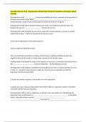
Complete fracture: break extends across the entire bone, dividing into two separate pieces
Incomplete fracture: bone only breaks partway across
Greenstick fracture: common in children. Bone is splintered on one side, but only bent on the other side
Classification of Fractures: Closed or simple fracture: The bone does not break through the skin
Open or compound fracture: Fragments of the broken bone break through skin, Open fractures have
three grades of severity
Grade I: least severe injury, with minimal skin damage
Grade II: moderately severe injury, with skin and muscle contusions (bruises)
Grade III: most severe injury (wound larger than 6 to 8 cm), with skin, muscle, blood vessel, and nerve
damage.
Stress fracture: Caused by either repeated or prolonged stress
Pathologic fracture: Occurs because of a pathologic condition in the bone, such as a tumor or disease
process, that causes a spontaneous break.
Cause and Risk Factors: Commonly caused by trauma to the bone, especially as a result of automobile
accidents and falls, Bone disease; e.g., bone cancer, can lead to a fracture, Hip fractures in older adults
usually from falls, Risk factors for hip fractures: osteoporosis, advanced age, white race, use of
psychotropic drugs, and female, In adults, ribs most commonly fractured, Fractures of the femur most
common in young and middle-aged adults, Hip and wrist fractures are most common in older adults.
Stage 4: ossification: Within 3 weeks to 6 months after the break, a permanent bone callus, known as
woven bone, forms. During this stage, the ends of the broken bone begin to knit.
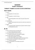
Complication - Fat Embolism: Fat globules released from marrow of broken bone into bloodstream, then
migrate to the lungs. They lodge in capillaries and obstruct blood flow. The fat particles break down into
fatty acids, which inflame the pulmonary blood vessels, leading to pulmonary edema. Common with
fractures of the long bones, multiple fractures, and severe trauma.
Complication – Shock: After fracture, a risk of excessive blood loss. Trauma may rupture local blood
vessels; internal organs may be punctured; results in internal bleeding. Loss of blood leads to shock,
evidenced by tachycardia; anxiety; pallor; and cool, clammy skin. Immobilizing fractures reduces risk of
hemorrhage. If severe external bleeding, external pressure should be applied, and medical assistance
summoned immediately.
Complication - Avascular Necrosis: A variety of factors can interfere with blood supply after a bone
injury. Once bone cells are deprived of oxygen and nutrients, they die and their cell walls collapse, Signs
and symptoms: Pain, instability, and decreased function in the affected area.
, Complication - Complex Regional Pain Syndrome Type 1 (CRPS—Type 1): Precipitated by a fracture or
other trauma. Symptoms:Severe pain at the injury site despite no detectable nerve damage, edema,
muscle spasm, stiffness, vasospasms, increased sweating, atrophy, contractions, and loss of bone mass,
Symptoms persist longer than expected with the type of injury suffered.
Reduction: The process of bringing the ends of the broken bone into proper alignment.
Closed Reduction or Manipulation: Nonsurgical realignment that returns bones to their previous
anatomic position. No surgical incision is made; however, general or local anesthesia is given. By using
traction, manual pressure, or a combination. After reduction of a fracture, x-ray taken and a cast usually
applied.
Casts, Splints, and Immobilizers: Hold the bone in alignment while allowing movement of other parts of
the body.Types of cast materials: plaster of Paris, fiberglass, thermoplastic resins, thermolabile plastic,
and polyester-cotton knit impregnated with polyurethane. Variety of materials used for
splints/immobilizers. Four main groups of casts: (1) upper extremity, (2) lower extremity, (3) cast brace,
and (4) body or spica cast.
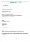
Traction: Skin traction: Buck’s traction For hip and knee contractures, muscle spasms, and alignment of
hip fractures. Weight used during skin traction should not be more than 5 to 10 pounds to prevent injury
to the skin
Skeletal traction: Provides a strong, steady, continuous pull and can be used for prolonged periods
Examples of skeletal traction are Gardner-Wells, Crutchfield, and Vinke tongs and a halo vest, in which
pins are inserted into the skull on either side.
Crutches: Increase mobility and assist with ambulation. Physical therapist measures patient for proper
fit and instructs in crutch-walking techniques. Nurse reinforces the instructions and evaluates whether
the crutches are being used properly. The type of gait used depends on the severity of the patient’s
disability and the patient’s physical condition, trunk strength, upper and lower extremity strength, and
balance. A properly fitted crutch should reach to three fingerbreadths below the axilla to avoid pressure
on the axilla and nerves when walking. Axillary pressure could result in temporary or permanent
numbness in the hands.
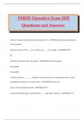
Colles Fracture: A break in the distal radius (wrist area). Colles fractures often occur in older adults,
particularly older women, when an outstretched hand is used to break a fall. The most common
complication is impaired circulation in the area resulting from edema. Medical diagnosis: Radiography.
Medical treatment: Closed reduction or manipulation of the bone and immobilization in either a splint
or a cast.
Amputation
Amputation: Can occur through a joint (between the bones) or through a bone itself. Disarticulation:
term used for an amputation through the joint. The general site of the amputation is described by the
joint nearest to it.
Introduction: Fracture is a break or disruption in the continuity of a bone
Complete fracture: break extends across the entire bone, dividing into two separate pieces
Incomplete fracture: bone only breaks partway across
Greenstick fracture: common in children. Bone is splintered on one side, but only bent on the other side
Classification of Fractures: Closed or simple fracture: The bone does not break through the skin
Open or compound fracture: Fragments of the broken bone break through skin, Open fractures have
three grades of severity
Grade I: least severe injury, with minimal skin damage
Grade II: moderately severe injury, with skin and muscle contusions (bruises)
Grade III: most severe injury (wound larger than 6 to 8 cm), with skin, muscle, blood vessel, and nerve
damage.
Stress fracture: Caused by either repeated or prolonged stress
Pathologic fracture: Occurs because of a pathologic condition in the bone, such as a tumor or disease
process, that causes a spontaneous break.
Cause and Risk Factors: Commonly caused by trauma to the bone, especially as a result of automobile
accidents and falls, Bone disease; e.g., bone cancer, can lead to a fracture, Hip fractures in older adults
usually from falls, Risk factors for hip fractures: osteoporosis, advanced age, white race, use of
psychotropic drugs, and female, In adults, ribs most commonly fractured, Fractures of the femur most
common in young and middle-aged adults, Hip and wrist fractures are most common in older adults.
Stage 4: ossification: Within 3 weeks to 6 months after the break, a permanent bone callus, known as
woven bone, forms. During this stage, the ends of the broken bone begin to knit.
Complication - Fat Embolism: Fat globules released from marrow of broken bone into bloodstream, then
migrate to the lungs. They lodge in capillaries and obstruct blood flow. The fat particles break down into
fatty acids, which inflame the pulmonary blood vessels, leading to pulmonary edema. Common with
fractures of the long bones, multiple fractures, and severe trauma.
Complication – Shock: After fracture, a risk of excessive blood loss. Trauma may rupture local blood
vessels; internal organs may be punctured; results in internal bleeding. Loss of blood leads to shock,
evidenced by tachycardia; anxiety; pallor; and cool, clammy skin. Immobilizing fractures reduces risk of
hemorrhage. If severe external bleeding, external pressure should be applied, and medical assistance
summoned immediately.
Complication - Avascular Necrosis: A variety of factors can interfere with blood supply after a bone
injury. Once bone cells are deprived of oxygen and nutrients, they die and their cell walls collapse, Signs
and symptoms: Pain, instability, and decreased function in the affected area.
, Complication - Complex Regional Pain Syndrome Type 1 (CRPS—Type 1): Precipitated by a fracture or
other trauma. Symptoms:Severe pain at the injury site despite no detectable nerve damage, edema,
muscle spasm, stiffness, vasospasms, increased sweating, atrophy, contractions, and loss of bone mass,
Symptoms persist longer than expected with the type of injury suffered.
Reduction: The process of bringing the ends of the broken bone into proper alignment.
Closed Reduction or Manipulation: Nonsurgical realignment that returns bones to their previous
anatomic position. No surgical incision is made; however, general or local anesthesia is given. By using
traction, manual pressure, or a combination. After reduction of a fracture, x-ray taken and a cast usually
applied.
Casts, Splints, and Immobilizers: Hold the bone in alignment while allowing movement of other parts of
the body.Types of cast materials: plaster of Paris, fiberglass, thermoplastic resins, thermolabile plastic,
and polyester-cotton knit impregnated with polyurethane. Variety of materials used for
splints/immobilizers. Four main groups of casts: (1) upper extremity, (2) lower extremity, (3) cast brace,
and (4) body or spica cast.
Traction: Skin traction: Buck’s traction For hip and knee contractures, muscle spasms, and alignment of
hip fractures. Weight used during skin traction should not be more than 5 to 10 pounds to prevent injury
to the skin
Skeletal traction: Provides a strong, steady, continuous pull and can be used for prolonged periods
Examples of skeletal traction are Gardner-Wells, Crutchfield, and Vinke tongs and a halo vest, in which
pins are inserted into the skull on either side.
Crutches: Increase mobility and assist with ambulation. Physical therapist measures patient for proper
fit and instructs in crutch-walking techniques. Nurse reinforces the instructions and evaluates whether
the crutches are being used properly. The type of gait used depends on the severity of the patient’s
disability and the patient’s physical condition, trunk strength, upper and lower extremity strength, and
balance. A properly fitted crutch should reach to three fingerbreadths below the axilla to avoid pressure
on the axilla and nerves when walking. Axillary pressure could result in temporary or permanent
numbness in the hands.
Colles Fracture: A break in the distal radius (wrist area). Colles fractures often occur in older adults,
particularly older women, when an outstretched hand is used to break a fall. The most common
complication is impaired circulation in the area resulting from edema. Medical diagnosis: Radiography.
Medical treatment: Closed reduction or manipulation of the bone and immobilization in either a splint
or a cast.
Amputation
Amputation: Can occur through a joint (between the bones) or through a bone itself. Disarticulation:
term used for an amputation through the joint. The general site of the amputation is described by the
joint nearest to it.