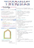
VERTEBRATE IMMUNE SYSTEM
The vertebrate immune system has two components: the innate and adaptive system. The innate
immunity consists of external barriers (skin, mucous membrane) and internal barriers that form the
first line of defence (phagocytes, natural killer cells, TLRs). The adaptive immunity is the internal
second line of defence (T-cells, B-cells, Ig), this response is much slower than the innate. These two
systems interact and cooperate. Major innate immune cell types are neutrophils (phagocytosis, ROS,
antimicrobial peptides), macrophages (phagocytosis, inflammatory mediator, antigen presentation,
ROS, cytokines), dendritic cells (phagocytosis, antigen presentation, costimulatory signals, interferon)
and natural killer cells (lysis of viral-infected cells, interferon, macrophage activation). Antigen
presenting cells can engulf proteins, break them down into peptides and present these on MHC
receptors, where they are recognised by T-cells. The innate immune response recognises structurally
conserved features of microorganisms, PAMPs. Examples of this in gram-negative bacteria are
lipopolysaccharides, flagellin and unmethylated DNA. For viruses PAMPs can be glycoproteins and
ssRNA or dsRNA. PAMPs alone are not enough to activate an entire immune response, DAMPs are
also needed.
PAMPs and DAMPs are recognised
by Pattern Recognition Receptors
(PRRs). Common PRRs are Toll-like
receptors (TLRs). TLRs contain an
extracellular leucine-rich repeats
domain that recognised pathogens
and an intracellular TIR domain
which recruits a signalling complex.
TLRs can be located in the plasma
membrane or on internal vesicles
(endosomes). There are multiple
pathways that can occur after
pattern recognition by TLRs, they all
lead to the expression of
inflammatory genes. Some of these
genes encode for cytokines that will
bind to their own receptor and
amplify the inflammation signal.
Another type of PRR are inflammasomes. They are cytosolic and typically consist of a sensor protein,
ASC and caspase-1. An example is the NLRP3 inflammasome. When NLRP3, which is an intracellular
PRR, is activated by stress signals (K+ and CL- efflux, Ca2+ influx, ROS production), it oligomerises into
a complex, with the leucine-rich repeats on the outside (to recognise signals). The adaptor protein
ASC is then recruited to the inside of the complex, when there is enough of this, an inactive form of
caspase-1 is recruited. The inflammasome is now formed and pro-caspase-1 cleaves itself to become
activated. Activated caspase-1 can cleave cytokines like IL and Gasdermin-D. Gasdermin-D creates
pores in the cell membrane to release cytokines, this will eventually lead to rupture and cell death.
There are two signals required for inflammasome activation and initiation of pyroptosis. The first
signal is often a TLR that leads to the transcription of inflammasome components and cytokine
precursors. The second signal often responds to DAMPs and triggers the assembly of the
inflammasome. This is the canonical pathway, there is also a non-canonical pathway that does not
require ASC. This pathway is triggered by the presence of LPS, which binds to caspase 4/5. It is also
, very reliant on interferons. The non-canonical pathway is not host-beneficial. This activation will also
lead to an efflux of K+, which will activate the canonical pathway.
Activation of the NLRP3 inflammasome can happen by various mycobacterial proteins and lipids.
PPE13 participates in the assembly of NLRP3 inflammasome complex by interacting with the leucine-
rich repeat domains of NLPR3. EST12 is a pyroptosis-inducing protein that works through NLRP3. The
Mtb lipoprotein LpqH activates NLRP3 via a mechanism that involves activation of the TLR-2 receptor.
The ESX-1 and ESX-5 secretion systems are also very important. EsxA is one of the main effectors
secreted by ESX-1 and it interacts with TLR-2 and TLR-4 receptors and stimulates the translocation of
other inflammatory components into the cytosol. TDB is a cell wall lipid that is used in tuberculosis
vaccines because it acts as a PAMP. Mtb is capable of inhibiting the AIM2 inflammasome activation,
most likely with the help of the ESX-1 secretion system. Inhibition of the NLRP3 inflammasome also
occurs, but this happens independent of ESX-1. It works through inhibition of the LPS induced K+
influx, which leads to a decrease in ROS. Furthermore, the serine hydrolase Hip1, inhibits the NLRP3
inflammasome activation by dampening the TLR2-dependent signalling. Nitric oxide also acts as a
negative regulator as it inhibits processing of IL-1b. Lastly, host OXSR1 inhibits K+ channels that are
responsible for the K+ efflux and Mtb upregulates this gene.
There are three types of cell death: apoptosis (regulated process, cell shrinks, no inflammation),
necrosis (passive process, cell swells and ruptures, inflammation) and pyroptosis (regulated process,
cell swells, pore formation, strong inflammation). Inflammasomes induce cell death through
pyroptosis. Pyroptosis is always mediated by Gasdermin and almost always by inflammasomes. The
non-canonical inflammasome pathway still leads to pyroptosis but does not require ASC.
IL-1β is highly inflammatory and thus
the pathway is very regulated. Its
production relies on two different
pathways: the inflammasome
pathway and another signalling
pathway involving for example PRRs,
TLRs or cytokine receptors. The
binding of a ligand to these second
receptors can then for example lead
to the production of NF-kB, which
leads to the transcription of related
genes. In some cases, IL-1b can be
produced independent of
inflammasomes. This can be through
neutrophil involvement or activation
of caspase-8. IL-1b is proposed to
have both a protective and damaging
role for the host in Mtb infection. The protective role includes increasing apoptosis and autophagy
signalling. It also supresses IFN-β (which increases the host susceptibility), this however works both
ways. Contrary to this, an increase in IL-1b seems to be related to more severe tuberculosis.
Monocytes can differentiate into various types of macrophages; this is called macrophage
polarization. The two extremes are M1 macrophages which are very aggressive, pro-inflammatory,
bactericidal and phagocytic and M2 macrophages, which are anti-inflammatory, matrix producing,
pro-angiogenesis and pro-wound healing. The first type responds to recognition of PAMPs and
inflammatory immune signalling and leads to the activation of pro-inflammatory pathways through