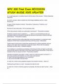
Exam 1 = feb 20 (cha 1, 3, 4, 5)
Chapter 3 Observing Microorganisms
Figure 3.2 Microscopes and magnification is a good summary about what microscopes is
used to observe certain things
Light Microscopy
Any kind of microscope that uses visible light to observe specimens
Types of light microscopy
o Compound light microscopy
o Darkfield microscopy
o Phase-contrast microscopy
o Differential interference contrast (DIC) microscopy (light comes from two
different sources)
o Fluorescence microscopy (used in short wavelength light e.g. lasers)
there are microbes that naturally fluoresce
Chronoforms- pigments we can attach to proteins that allows fluorescent
visualization
o Confocal microscopy (takes pictures of different planes)
The longer the wavelength → lesser resolution
Scanning Acoustic Microscopy
Measures sound waves that are reflected back from a specimen
Used to study cells attached to surfaces
Resolution of 1 m
Electron Microscopy
no light in this microscope
uses bouncing off of electrons, which is color enhanced by computer)
Transmission TEM
o 10,000 to 10,000,000x; resolution 10pm
Scanning (SEM)
o 1000 – 500,000x; resolution 10 nm
Scanning Microscopy
Scanning Tunneling
Atomic Force
Scanned-Probe
No lens
Figure 3.12a Gram Staining
1) Application of crystal violet (purple dye that goes into all the cells)
2) Application of iodine (mordant, helps make the purple dye stay)
3) Alcohol wash (decolorization)
4) Application of safranin (counter stain) 6
Gram positive stay purple, their cell walls collapse and stay purple
Gram negative stay pink
Differential Stains
Gram stain
o Gram-positive bacteria tend to be killed by penicillin + detergents
Purple = gram positive
, o Gram-negative bacteria are more resistant to antibiotics
Red = gram negative
Acid-fast stain
o Stained waxy cell wall is not decolorized by acid-alcohol
o Mycobacterium, Nocardia
o Red = acid-fast
Special Stains
Used to distinguish parts of microorganisms
o Capsule stain- negative staining
o Endospore stain- Schaeffer Fulton malachite green + heat; safe
o Flagella stain - mordant + carbolfuchsin
Chapter 4: Functional Anatomy of Prokaryotic and Eukaryotic Cells
Comparing Prokaryotic and Eukaryotic Cells: An Overview
Prokaryotes
o One circular chromosome not in a membrane
o No histones
o No organelles
o Bacteria: peptidoglycan cells walls
o Archaea: pseudomurein cell walls
o Divided by binary fission
Eukaryote
o Paired chromosomes, in nuclear membrane
o Histones
o Organelles
o Polysaccharide cell walls, when present
o Divides by mitosis
Basic Shapes
Bacillus (rod shaped)
o Both a genus (Italicized) and a shape
Coccus (spherical)
Spiral
o Spirillum
o Vibrio
o Spirochete
Monomorphic- one shape
Pleomorphic- multiple shapes
The Size, Shape, and Arrangement of Bacterial Cells (3 of 4)
Pairs: diplococci, diplobacilli
Clusters: staphylococci
Chains: streptococci, streptobacilli
Groups of Four: tetrads
Cubelike groups of eight: sarcinae
Figure 4.6 The Structure of a Prokaryotic Cell
Glycocalyx
Outside cell wall