Chapter 13 - The Molecular Basis of Inheritance
In April 1953, James Watson and Francis Crick shook the scientific world with an elegant double-helical
model for the three-dimensional structure of DNA.
DNA replication, also known as DNA synthesis, is the process by which a DNA molecule is copied.
13.1 DNA is the genetic material
In 1928, a British medical officer named Frederick Griffith was trying to develop a vaccine against
pneumonia. He was studying Streptococcus pneumoniae, a bacterium that causes pneumonia in mammals.
Griffith had two strains of the bacterium, one pathogenic (disease-causing) and one nonpathogenic
(harmless). He was surprised to find that when he killed the pathogenic bacteria with heat and then mixed
the cell remains with living bacteria of the nonpathogenic strain, some of the living cells became pathogenic.
This newly acquired trait of pathogenicity was inherited by all the descendants of the transformed bacteria.
Clearly, some chemical component of the dead pathogenic cells caused this heritable change. Griffith called
the phenomenon transformation, now defined as a change in genotype and phenotype due to the
assimilation of external DNA by a cell.
Additional evidence for DNA as the genetic material came from studies of viruses that infect bacteria. These
viruses are called bacteriophages or phages.
A virus is little more than DNA (or RNA) enclosed by a protective coat, which is often simply protein. To
produce more viruses, a virus must infect a cell and take over the cell's metabolic machinery.
In 1952, Alfred Hershey and Martha Chase performed experiments showing that DNA is the genetic material
of a phage known as T2. T2 infects E. coli, a bacterium that normally lives in the intestines of mammals and is
a model organism for molecular biologists.
In their experiment, they used a radioactive isotope of sulfur to tag proteins in one batch of T2 and a
radioactive isotope of phosphorus to tag DNA in a second batch. Because proteins, but not DNA, contain
sulfur, radioactive sulfur atoms were incorporated only into the protein of the phage. In conclusion, phage
DNA entered bacterial cells, but phage proteins did not.
Chargaff's rules: (1) the base composition varies between species, and (2) within a species, the numbers of A
and T bases are roughly equal and the numbers of G and C bases are roughly equal.
Double helix is the form of native DNA, referring to its two adjacent antiparallel polynucleotide strands
wound around an imaginary axis into a spiral shape.
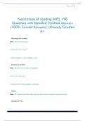
In this model, the two sugar-phosphate backbones are antiparallel; their subunits run in opposite directions.
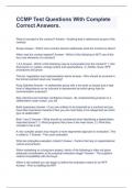
Adenine and guanine are purines, nitrogenous bases with two organic rings, while cytosine and thymine are
nitrogenous bases called pyrimidines, which have a single ring.
Thus, purines are about twice as wide as pyrimidines. A purine-purine pair
is too wide and a pyrimidine-pyrimidine pair too narrow to account for the
2nm diameter of the double helix.
Each base has chemical side groups that can form hydrogen bonds with its
appropriate partner: adenine forms two hydrogen bonds with thymine and
one thymine; guanine forms three hydrogen bonds with cytosine and only
cytosine.
13.2 Many proteins work together in DNA replication and repair
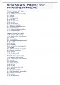
Watson and Crick's model predicts that when a double helix replicates,
each of the two daughter molecules will have one old strand, from the
parental molecule, and one newly made strand. This semiconservative
model can be distinguished from a conservative model of replication, in
which the two paternal strands somehow come back together after the
process. In yet a third model, called the dispersive model, all four strands
of DNA following replication have a mixture of old and new DNA.
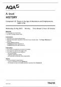
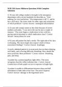
, The replication of a DNA molecule begins at particular sites
called origins of replication, short stretches of DNA having a
specific sequence of nucleotides.
Proteins that initiate DNA replication recognize this
sequence and attach to the DNA, separating the two strands
and opening up a replication bubble. At each end of the
bubble is a replication fork, a Y-shaped region where the
parental strands of DNA are being unwound.
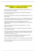
Helicases are enzymes that untwist the double helix at the
replication forks, separating the two paternal strands and
making them available as template strands.
After the paternal strands separate, single-strand binding
proteins bind to the unpaired DNA strands, keeping them for
re-pairing. The untwisting of the double helix causes tighter
twisting and strain ahead of the replication fork.
Topoisomerase helps relieve this strain by breaking,
swiveling, and rejoining DNA strands.
The initial nucleotide chain that is produced during DNA
synthesis is actually a short stretch of RNA, not DNA. This
RNA chain is called a primer and is synthesized by the
enzyme primase. Primase starts a complementary RNA
chain from a single RNA nucleotide, adding RNA nucleotides
In April 1953, James Watson and Francis Crick shook the scientific world with an elegant double-helical
model for the three-dimensional structure of DNA.
DNA replication, also known as DNA synthesis, is the process by which a DNA molecule is copied.
13.1 DNA is the genetic material
In 1928, a British medical officer named Frederick Griffith was trying to develop a vaccine against
pneumonia. He was studying Streptococcus pneumoniae, a bacterium that causes pneumonia in mammals.
Griffith had two strains of the bacterium, one pathogenic (disease-causing) and one nonpathogenic
(harmless). He was surprised to find that when he killed the pathogenic bacteria with heat and then mixed
the cell remains with living bacteria of the nonpathogenic strain, some of the living cells became pathogenic.
This newly acquired trait of pathogenicity was inherited by all the descendants of the transformed bacteria.
Clearly, some chemical component of the dead pathogenic cells caused this heritable change. Griffith called
the phenomenon transformation, now defined as a change in genotype and phenotype due to the
assimilation of external DNA by a cell.
Additional evidence for DNA as the genetic material came from studies of viruses that infect bacteria. These
viruses are called bacteriophages or phages.
A virus is little more than DNA (or RNA) enclosed by a protective coat, which is often simply protein. To
produce more viruses, a virus must infect a cell and take over the cell's metabolic machinery.
In 1952, Alfred Hershey and Martha Chase performed experiments showing that DNA is the genetic material
of a phage known as T2. T2 infects E. coli, a bacterium that normally lives in the intestines of mammals and is
a model organism for molecular biologists.
In their experiment, they used a radioactive isotope of sulfur to tag proteins in one batch of T2 and a
radioactive isotope of phosphorus to tag DNA in a second batch. Because proteins, but not DNA, contain
sulfur, radioactive sulfur atoms were incorporated only into the protein of the phage. In conclusion, phage
DNA entered bacterial cells, but phage proteins did not.
Chargaff's rules: (1) the base composition varies between species, and (2) within a species, the numbers of A
and T bases are roughly equal and the numbers of G and C bases are roughly equal.
Double helix is the form of native DNA, referring to its two adjacent antiparallel polynucleotide strands
wound around an imaginary axis into a spiral shape.
In this model, the two sugar-phosphate backbones are antiparallel; their subunits run in opposite directions.
Adenine and guanine are purines, nitrogenous bases with two organic rings, while cytosine and thymine are
nitrogenous bases called pyrimidines, which have a single ring.
Thus, purines are about twice as wide as pyrimidines. A purine-purine pair
is too wide and a pyrimidine-pyrimidine pair too narrow to account for the
2nm diameter of the double helix.
Each base has chemical side groups that can form hydrogen bonds with its
appropriate partner: adenine forms two hydrogen bonds with thymine and
one thymine; guanine forms three hydrogen bonds with cytosine and only
cytosine.
13.2 Many proteins work together in DNA replication and repair
Watson and Crick's model predicts that when a double helix replicates,
each of the two daughter molecules will have one old strand, from the
parental molecule, and one newly made strand. This semiconservative
model can be distinguished from a conservative model of replication, in
which the two paternal strands somehow come back together after the
process. In yet a third model, called the dispersive model, all four strands
of DNA following replication have a mixture of old and new DNA.
, The replication of a DNA molecule begins at particular sites
called origins of replication, short stretches of DNA having a
specific sequence of nucleotides.
Proteins that initiate DNA replication recognize this
sequence and attach to the DNA, separating the two strands
and opening up a replication bubble. At each end of the
bubble is a replication fork, a Y-shaped region where the
parental strands of DNA are being unwound.
Helicases are enzymes that untwist the double helix at the
replication forks, separating the two paternal strands and
making them available as template strands.
After the paternal strands separate, single-strand binding
proteins bind to the unpaired DNA strands, keeping them for
re-pairing. The untwisting of the double helix causes tighter
twisting and strain ahead of the replication fork.
Topoisomerase helps relieve this strain by breaking,
swiveling, and rejoining DNA strands.
The initial nucleotide chain that is produced during DNA
synthesis is actually a short stretch of RNA, not DNA. This
RNA chain is called a primer and is synthesized by the
enzyme primase. Primase starts a complementary RNA
chain from a single RNA nucleotide, adding RNA nucleotides