CH 24
1. overview
- immune system: physiological system whose primary job is to protect the body from
damage
- body’s ability to protect itself is known as immunity
- nonself: includes bacteria; virus; … and other disease causing pathogens
- body’s first line of defense against pathogens includes physical; chemical;
mechanical barriers (skin); tears; mucus and stomach acid
→ attempt to keep pathogens from entering ECF
but if they evade them the body initiates an immediate response: 4 steps
➢ detection and identification of the pathogen
➢ communication with other immune cells to rally an organized response
➢ recruitment of assistance and coordination of the response among participants
➢ destruction / suppression of pathogen
- substances that trigger the body’s immune response: immunogens
- immunogens that react with products of the immune response are known as antigens
- inter immune response is carried out by the leukocytes, dependant on cell-cell
communication
- chemical communication includes substances released by damaged or dying cells as
well as cytokines (protein signal molecules released by one cell that affect the growth
or activity of another cell)
- immune system is also the primary user of contact dependent signaling: when
surface receptors on one cell recognize and bind to surface receptors on another cell
- internal immune response can be divided into 2 phases
➢ a rapid innate response
➢ slower adaptive response
1.1 innate immunity
- present from birth
- body’s immediate response to invasion
- not specific to a pathogen
- inflammation: visible on skin when red; warm; swollen → sign of innate immunity
- a innate response to pathogen in not remembered by the immune system and must
be triggered anew with each exposure
- cell responsible for the rapid response: circulating and stationary leukocytes that are
programmed to respond to a broad range of material
vb. phagocytes identify bacteria as pathogen; they ingest it via phagocytosis and digest it
- cells that display pathogen this way are called antigen presenting cells (APCs)
1.2 adaptive immunity
- directed at particular invaders of the body’s specific immune response
- steps needed to launch a specific immune response may take days to week
- re exposure: certain immune cells called memory cells remember the prior exposure;
react rapidly
, - it can be divided into 2 categories
➢ cell mediated immunity: B en T lymphocyten
➢ antibody mediated immunity: antibodies
1.2.1 cell mediated immunity
= acquired immunity
- requires contact dependent signaling between immune cell and receptors on its
target cell
1.2.2 antibody mediated immunity
= humorale immuniteit
- uses antibodies (proteins secreted by immune cells to carry out immunes response)
- antibodies binds to foreign substances to disable them or make more visible for cells
of immune system
- humoral: referring to the blood; comes from school of medicine which classified
body’s fluid into four humors: blood; phlegm; black bile; yellow bile
★ the innate and adaptive immunity overlap; we described them as separate but in
reality they are interconnected parts of single process
★ innate response: more rapid response; not target specific invader; reinforced by
antigen specific adaptive response
★ not all pathogens can be destroyed by immune system; sometimes damage the
control and keep invader from spreading
vb. tbc: hides inside cells in the lung; malaria parasites hide inside liver…
immune system serves three major functions
➢ it tries to recognize and remove abnormal self cells: created when normal cell growth
goes wrong
➢ it removes dead or damaged cells: as well as old RBC; scavenger cells of immune
system patrol the EC compartment; digesting dead or dying cells
➢ it protects the body from disease causing pathogens: microbes act as pathogens
include bacteria; virus: larger pathogens include multicellular parasites
2. anatomy of the immune system
2 anatomical components
➢ lymphoid tissues
➢ cells responsible for immune response: positioned wherever pathogens are likely to
enter the body
2.1 lymphoid tissues
- found all over the body
- primary lymphoid tissues are the thymus (produces T lymphocytes) gland and bone
marrow (produces RBC)
- secondary lymphoid tissues: mature immune cells interact with pathogens and initiate
a response
➢ encapsulated tissues: spleen and lymph nodes: monitor EC compartment for foreign
invaders; phagocytic cells in spleen trap and remove aging RBC; lymph nodes part of
lymphatic circulation (capillaries of CVS; BP creates net flow of fluid out of blood
, capillaries into interstitial space); filtered fluid: picked up by lymph capillaries and
passes through encapsulated lymph nodes on its way back to the heart
- once microbes in lymphatic circulation; immune cells in lymph nodes try to capture
them to prevent their spread
➢ unencapsulated tissues: diffuse lymphoid tissues; appear in other organs and
tissues; include cells in the skin; tonsils (De keelamandelen of tonsillen bevinden zich
in de keelholte en werken als een soort filter die binnenkomende ziektekiemen
kunnen bestrijden); cells with mucosal surfaces
- mucosa associated lymphoid tissue (MALT)
- subgroups of MALT include the gut associated lymphoid tissue (GALT), lies under the
epithelium of esophagus and intestines
- each location: immune cells positioned to intercept invading pathogens before the
general circulation
WBC (leukocyten):
- primary cell type responsible for immune responses
- much larger than RBC
- not very numerous in the circulation (5 million RBC; 7000 WBC)
- circulate in blood: they also leave the capillaries and function extravascularly
- some types of WBC live out in the tissue for months; others only days
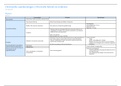
- divided into 6 basic types
➔ basophils: in the blood and related to mast cells in tissues
➔ eosinophils
➔ neutrophils
➔ monocytes and their derivative macrophages
➔ dendritic cells
➔ lymphocytes and their derivative plasma cells
- dendritic and mast cells not usually found in the blood; often excluded of leukocytes
in blood
- leukocytes can be distinguished from one another in stained tissue samples by the
shape and size of the nucleus
immune cell names
- one morphological group of leukocytes is the granulocytes
→ whose plasma contains granules
- granulocytes includes: basophils; eosinophils; neutrophils
★ basophil granules: dark blue with basic dye
★ eosinophil granules: dark pink with acidic dye eosin (dawn)
★ neutrophil granules: not stain darkly with blood and is neutral
→ they all release it granules by exocytosis (degranulation)
- 2 function group of leukocytes
➢ phagocytes: ingest material from ECF using large vesicle; include neutrophils;
macrophages; dendritic cells
➢ antigen presenting cell: APCs: ability to du-isplay bits of antigen on their surface as a
signal to other immune cells
→ primary APC: are the macrophages and dendritic cells
1. overview
- immune system: physiological system whose primary job is to protect the body from
damage
- body’s ability to protect itself is known as immunity
- nonself: includes bacteria; virus; … and other disease causing pathogens
- body’s first line of defense against pathogens includes physical; chemical;
mechanical barriers (skin); tears; mucus and stomach acid
→ attempt to keep pathogens from entering ECF
but if they evade them the body initiates an immediate response: 4 steps
➢ detection and identification of the pathogen
➢ communication with other immune cells to rally an organized response
➢ recruitment of assistance and coordination of the response among participants
➢ destruction / suppression of pathogen
- substances that trigger the body’s immune response: immunogens
- immunogens that react with products of the immune response are known as antigens
- inter immune response is carried out by the leukocytes, dependant on cell-cell
communication
- chemical communication includes substances released by damaged or dying cells as
well as cytokines (protein signal molecules released by one cell that affect the growth
or activity of another cell)
- immune system is also the primary user of contact dependent signaling: when
surface receptors on one cell recognize and bind to surface receptors on another cell
- internal immune response can be divided into 2 phases
➢ a rapid innate response
➢ slower adaptive response
1.1 innate immunity
- present from birth
- body’s immediate response to invasion
- not specific to a pathogen
- inflammation: visible on skin when red; warm; swollen → sign of innate immunity
- a innate response to pathogen in not remembered by the immune system and must
be triggered anew with each exposure
- cell responsible for the rapid response: circulating and stationary leukocytes that are
programmed to respond to a broad range of material
vb. phagocytes identify bacteria as pathogen; they ingest it via phagocytosis and digest it
- cells that display pathogen this way are called antigen presenting cells (APCs)
1.2 adaptive immunity
- directed at particular invaders of the body’s specific immune response
- steps needed to launch a specific immune response may take days to week
- re exposure: certain immune cells called memory cells remember the prior exposure;
react rapidly
, - it can be divided into 2 categories
➢ cell mediated immunity: B en T lymphocyten
➢ antibody mediated immunity: antibodies
1.2.1 cell mediated immunity
= acquired immunity
- requires contact dependent signaling between immune cell and receptors on its
target cell
1.2.2 antibody mediated immunity
= humorale immuniteit
- uses antibodies (proteins secreted by immune cells to carry out immunes response)
- antibodies binds to foreign substances to disable them or make more visible for cells
of immune system
- humoral: referring to the blood; comes from school of medicine which classified
body’s fluid into four humors: blood; phlegm; black bile; yellow bile
★ the innate and adaptive immunity overlap; we described them as separate but in
reality they are interconnected parts of single process
★ innate response: more rapid response; not target specific invader; reinforced by
antigen specific adaptive response
★ not all pathogens can be destroyed by immune system; sometimes damage the
control and keep invader from spreading
vb. tbc: hides inside cells in the lung; malaria parasites hide inside liver…
immune system serves three major functions
➢ it tries to recognize and remove abnormal self cells: created when normal cell growth
goes wrong
➢ it removes dead or damaged cells: as well as old RBC; scavenger cells of immune
system patrol the EC compartment; digesting dead or dying cells
➢ it protects the body from disease causing pathogens: microbes act as pathogens
include bacteria; virus: larger pathogens include multicellular parasites
2. anatomy of the immune system
2 anatomical components
➢ lymphoid tissues
➢ cells responsible for immune response: positioned wherever pathogens are likely to
enter the body
2.1 lymphoid tissues
- found all over the body
- primary lymphoid tissues are the thymus (produces T lymphocytes) gland and bone
marrow (produces RBC)
- secondary lymphoid tissues: mature immune cells interact with pathogens and initiate
a response
➢ encapsulated tissues: spleen and lymph nodes: monitor EC compartment for foreign
invaders; phagocytic cells in spleen trap and remove aging RBC; lymph nodes part of
lymphatic circulation (capillaries of CVS; BP creates net flow of fluid out of blood
, capillaries into interstitial space); filtered fluid: picked up by lymph capillaries and
passes through encapsulated lymph nodes on its way back to the heart
- once microbes in lymphatic circulation; immune cells in lymph nodes try to capture
them to prevent their spread
➢ unencapsulated tissues: diffuse lymphoid tissues; appear in other organs and
tissues; include cells in the skin; tonsils (De keelamandelen of tonsillen bevinden zich
in de keelholte en werken als een soort filter die binnenkomende ziektekiemen
kunnen bestrijden); cells with mucosal surfaces
- mucosa associated lymphoid tissue (MALT)
- subgroups of MALT include the gut associated lymphoid tissue (GALT), lies under the
epithelium of esophagus and intestines
- each location: immune cells positioned to intercept invading pathogens before the
general circulation
WBC (leukocyten):
- primary cell type responsible for immune responses
- much larger than RBC
- not very numerous in the circulation (5 million RBC; 7000 WBC)
- circulate in blood: they also leave the capillaries and function extravascularly
- some types of WBC live out in the tissue for months; others only days
- divided into 6 basic types
➔ basophils: in the blood and related to mast cells in tissues
➔ eosinophils
➔ neutrophils
➔ monocytes and their derivative macrophages
➔ dendritic cells
➔ lymphocytes and their derivative plasma cells
- dendritic and mast cells not usually found in the blood; often excluded of leukocytes
in blood
- leukocytes can be distinguished from one another in stained tissue samples by the
shape and size of the nucleus
immune cell names
- one morphological group of leukocytes is the granulocytes
→ whose plasma contains granules
- granulocytes includes: basophils; eosinophils; neutrophils
★ basophil granules: dark blue with basic dye
★ eosinophil granules: dark pink with acidic dye eosin (dawn)
★ neutrophil granules: not stain darkly with blood and is neutral
→ they all release it granules by exocytosis (degranulation)
- 2 function group of leukocytes
➢ phagocytes: ingest material from ECF using large vesicle; include neutrophils;
macrophages; dendritic cells
➢ antigen presenting cell: APCs: ability to du-isplay bits of antigen on their surface as a
signal to other immune cells
→ primary APC: are the macrophages and dendritic cells