Functional Magnetic Resonance Imaging (fMRI)
- Allows studying the human brain in action
- Non-invasive measurement of brain activity.
- Appealing balance between:
- Temporal resolution: Seconds
- Spatial resolution: Millimeter
- Coverage: Whole-brain
- Imaging analysis skills are a sought after also in other disciplines
- Inherently interdisciplinary field
Why not fMRI: Common criticism:
- Indirect (blood flow related) measure of neural activity: Hemodynamic coupling is well
established, and at least it measures everything, not just the effect of neuronal
spikes.
- Limited resolution (e.g. no single cells, no action potentials)
- Constrained set of experiments (e.g. head motion is not possible)
- MRI signals are noisy (e.g., often low signal-to-noise ratios)
- Analytical challenges (e.g. autocorrelations)
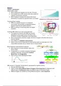
1. Main magnet
Creates a strong magnetic field
(7 Tesla = 140.000x earth's magnetic field)
2. Radiofrequency (RF) coil
Transmits & receives radio frequency waves
3. Gradient coils
Create additional magnetic fields whose
strength varies along XYZ dimensions
(important for localization the signal)
4. Patient table
Moves the patient in and out
5. Computer system
Controls the scanner from another room
Field strength: 1 Tesla is equal to 20.000 Earth’s magnetic field
Typical hospital scanner is 1.5-3 Tesla
Research scanners are between 3 and 11 Tesla
Different images are created by running different programs called MRI sequences.
MR-physics and data acquisition, when there is no net-magnetization, every
proton points in a random axis in a random phase.
- Longitudinal magnetization (T1): Axis aligned to B0, random phase:
strong magnetic field (B0)
- Transverse magnetization (T2): Axis flipped orthogonal to B0, protons
now phase aligned → strong magnetic field (B0) → Radiofrequency pulse
Phase coherence gets lost
,Protons resonate if the RF-pulse frequency matches their precession frequency (i.e. they
take on energy)
T1-weighted image T2-weighted image
→ Structural images → Functional images
Higher resolution (±1 mm at 3T) Lower resolution (±2mm at 3T)
High contrast, fewer artifacts Susceptible to blood oxygenation (T2*)
Voxel = Each pixel corresponds to a three-dimensional square or rectangular chunk of brain
tissue called a volume element
Time between images = Repetition time (TR)
Preprocessing:
1. Motion correction:
Problem → Head movements shift voxels
Solution → Realignment: Images are rotated & moved until they algin
2. Unwarping:
Problem → Recorded image is often distorted
Solution → Map and correct magnetic field distortions: Images are corrected for
warping
3. Slice time correction:
Problem → Slices acquired sequentially
Solution → Shifting signal depending on acquisition time, Resampling at interpolated
time points. Becomes more important the longer the TR.
4. Coregistration:
Problem → Structural and functional images need to be aligned but differ in contrast
& artifacts
Solution → Maximize mutual information, aligning images with an algorithm handling
different contrasts (unlike motion correction)
5. Normalization:
Problem → Anatomical differences between subjects, coordinates are not
comparable.
Solution → Convert images into common space, Warping all images of all
participants such that they align with the same template brain.
6. Spatial smoothing:
Problem → Weak signals & residual anatomical differences
Solutions → Smoothing images suppresses noise and increases statistical power.
, Each voxel is replaced by a Gaussian-weighted average of itself and its neighbors.
con → Very spatially specific activations, for example in small midbrain nuclei,
disappear
Data analysis
- Blood Oxygenation Level Dependent (BOLD) Signal
- MRI → Stimulus → Hemodynamic response function (HRF)
- Stimulus Onsets + HRF → Predicted BOLD signal
General Linear Model (GLM)
- Hot colors show voxels whose time series could be well predicted based on the
experiment
fMRI experiments
Task-based vs. Task-free approaches in Cognitive Neuroscience
Ways to measure behavior & physiological signals in fMRI
- Button boxes & joysticks → Basic behavioral responses
- Eye tracking → Gaze behavior & pupil size
- Microphones → Speech
- Pneumograph belts → Breathing
- Pulse oximeter → Blood oxygenation, heart rate
- Experimental considerations → e.g. video games
Ways to present stimuli
- Screen + Mirror
- Goggles & Headphones: 3D stimuli, virtual reality
- Vibration devices: tactile stimuli, touch
- Galvanic stimulator: vestibular stimuli, perceived movement
- Gustometers & odor stimulators: taste, smells or flavors
- Brain stimulation: magnetic or electrical
- Allows studying the human brain in action
- Non-invasive measurement of brain activity.
- Appealing balance between:
- Temporal resolution: Seconds
- Spatial resolution: Millimeter
- Coverage: Whole-brain
- Imaging analysis skills are a sought after also in other disciplines
- Inherently interdisciplinary field
Why not fMRI: Common criticism:
- Indirect (blood flow related) measure of neural activity: Hemodynamic coupling is well
established, and at least it measures everything, not just the effect of neuronal
spikes.
- Limited resolution (e.g. no single cells, no action potentials)
- Constrained set of experiments (e.g. head motion is not possible)
- MRI signals are noisy (e.g., often low signal-to-noise ratios)
- Analytical challenges (e.g. autocorrelations)
1. Main magnet
Creates a strong magnetic field
(7 Tesla = 140.000x earth's magnetic field)
2. Radiofrequency (RF) coil
Transmits & receives radio frequency waves
3. Gradient coils
Create additional magnetic fields whose
strength varies along XYZ dimensions
(important for localization the signal)
4. Patient table
Moves the patient in and out
5. Computer system
Controls the scanner from another room
Field strength: 1 Tesla is equal to 20.000 Earth’s magnetic field
Typical hospital scanner is 1.5-3 Tesla
Research scanners are between 3 and 11 Tesla
Different images are created by running different programs called MRI sequences.
MR-physics and data acquisition, when there is no net-magnetization, every
proton points in a random axis in a random phase.
- Longitudinal magnetization (T1): Axis aligned to B0, random phase:
strong magnetic field (B0)
- Transverse magnetization (T2): Axis flipped orthogonal to B0, protons
now phase aligned → strong magnetic field (B0) → Radiofrequency pulse
Phase coherence gets lost
,Protons resonate if the RF-pulse frequency matches their precession frequency (i.e. they
take on energy)
T1-weighted image T2-weighted image
→ Structural images → Functional images
Higher resolution (±1 mm at 3T) Lower resolution (±2mm at 3T)
High contrast, fewer artifacts Susceptible to blood oxygenation (T2*)
Voxel = Each pixel corresponds to a three-dimensional square or rectangular chunk of brain
tissue called a volume element
Time between images = Repetition time (TR)
Preprocessing:
1. Motion correction:
Problem → Head movements shift voxels
Solution → Realignment: Images are rotated & moved until they algin
2. Unwarping:
Problem → Recorded image is often distorted
Solution → Map and correct magnetic field distortions: Images are corrected for
warping
3. Slice time correction:
Problem → Slices acquired sequentially
Solution → Shifting signal depending on acquisition time, Resampling at interpolated
time points. Becomes more important the longer the TR.
4. Coregistration:
Problem → Structural and functional images need to be aligned but differ in contrast
& artifacts
Solution → Maximize mutual information, aligning images with an algorithm handling
different contrasts (unlike motion correction)
5. Normalization:
Problem → Anatomical differences between subjects, coordinates are not
comparable.
Solution → Convert images into common space, Warping all images of all
participants such that they align with the same template brain.
6. Spatial smoothing:
Problem → Weak signals & residual anatomical differences
Solutions → Smoothing images suppresses noise and increases statistical power.
, Each voxel is replaced by a Gaussian-weighted average of itself and its neighbors.
con → Very spatially specific activations, for example in small midbrain nuclei,
disappear
Data analysis
- Blood Oxygenation Level Dependent (BOLD) Signal
- MRI → Stimulus → Hemodynamic response function (HRF)
- Stimulus Onsets + HRF → Predicted BOLD signal
General Linear Model (GLM)
- Hot colors show voxels whose time series could be well predicted based on the
experiment
fMRI experiments
Task-based vs. Task-free approaches in Cognitive Neuroscience
Ways to measure behavior & physiological signals in fMRI
- Button boxes & joysticks → Basic behavioral responses
- Eye tracking → Gaze behavior & pupil size
- Microphones → Speech
- Pneumograph belts → Breathing
- Pulse oximeter → Blood oxygenation, heart rate
- Experimental considerations → e.g. video games
Ways to present stimuli
- Screen + Mirror
- Goggles & Headphones: 3D stimuli, virtual reality
- Vibration devices: tactile stimuli, touch
- Galvanic stimulator: vestibular stimuli, perceived movement
- Gustometers & odor stimulators: taste, smells or flavors
- Brain stimulation: magnetic or electrical