Methods
🧠perception- how we recognize things in the world
From Sensation to Perception
🧠sensation- the physical process of taking the physicalenergy in our environment and
converting into neural signals that the brain can understand
🧠
perception- the brain’s interpretation the neuralsignals in order to understand what’s going
on in the environment
🧠
stimuli → sensation → perception
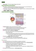
Gross Anatomy of the Eye
🧠photoreceptorsreside at the back of the eye (retina)
○ captures the light from the environment and make sense of it
○ sends information perceived to the brain for processing
🧠
thelensrefracts the image to the photoreceptors
🧠
the eye has a spot considered theblindspot
○ part of the retina that don’t have any photoreceptors
○ AKA the optic nerve
○ located at the back of the eye where the cells exit the eye to feed the brain information
■ the left eye fills in blanks in the environment of the visual right field, vice versa
for the right eye and the left visual field
🧠
bipolar andganglion cellsrepresent diff info
○ more of these cells = more info being represented
🧠
light has to pass through the translucent cells found in the middle layer
Visual deficits
🧠
can be either be because of eye problems or problems in the brain
🧠
a problem with the eye that can cause visual deficit could be because ofretina detaching
○ lindness can be caused if it becomes fully detached
b
○ associated w/ medical conditions [i.e. high BP, diabetes, etc.)
○ slowly starts peeling off from the back of the eye
○ the longer you wait to fix the problem causes it to detach more, making it harder to
fix/reattach
🧠
solution for retina detachment =retinal implant device
, m
○ imics what the retina would do
○ invasive process but still game changing for this visual problem
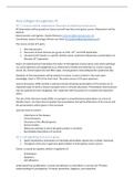
Primary Projection Pathways of Visual System
🧠
each eye contributes to each side of the visual fields
🧠
theoptic chiasmis where the optic nerves cross
○ peripheral vision crosses here
🧠
thepituitary glandsits on top of the optic chiasm
○ when the PG swells or becomes cancerous, it inhibits the optic chiasm from
processing the side fields
■ this causestunnel vision
■ inhibits peripheral vision
🧠
visual impairment can be caused either at the eye level or at a neurological level
○ neurological level =damage to one of the brain structuresa part of the visual pathways
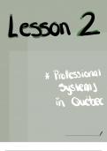
Blind Spots Caused by Brain Injury
🧠retinotopic mapping= a map created by the brain toreplicate the world being perceived
○ onto the primary visual cortex
🧠
injury to the retinotopic map causesscotomathatcorresponds to that location
○ scotoma = a blindspot or visual field abnormality that can occur in either one or both
eyes
🧠
fovea is the center of the retinotopic mapping, captures what the individual is directly
looking at
🧠
the fovea is split into 12 parts that correspond to an area of the visual
cortex map
🧠
damage to the entire left occipital lobe impacts the right visual field
🧠
when there are visual impairments that stem from damage to the
brain the blindness is categorized ascortical blindness
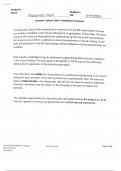
Congenital Blindness
🧠
blind from birth
🧠
study shows that individuals that are born blind and learn braille later in life show activation
in the occipital lobe
🧠
the evidence shows that the occipital lobe is rewired/remapped to help with the sense of
touch
○ this rewiring is able to occur due to neuroplasticity
🧠
the ‘touch lobe’ still works the way it usually does, just with a little more help
🧠perception- how we recognize things in the world
From Sensation to Perception
🧠sensation- the physical process of taking the physicalenergy in our environment and
converting into neural signals that the brain can understand
🧠
perception- the brain’s interpretation the neuralsignals in order to understand what’s going
on in the environment
🧠
stimuli → sensation → perception
Gross Anatomy of the Eye
🧠photoreceptorsreside at the back of the eye (retina)
○ captures the light from the environment and make sense of it
○ sends information perceived to the brain for processing
🧠
thelensrefracts the image to the photoreceptors
🧠
the eye has a spot considered theblindspot
○ part of the retina that don’t have any photoreceptors
○ AKA the optic nerve
○ located at the back of the eye where the cells exit the eye to feed the brain information
■ the left eye fills in blanks in the environment of the visual right field, vice versa
for the right eye and the left visual field
🧠
bipolar andganglion cellsrepresent diff info
○ more of these cells = more info being represented
🧠
light has to pass through the translucent cells found in the middle layer
Visual deficits
🧠
can be either be because of eye problems or problems in the brain
🧠
a problem with the eye that can cause visual deficit could be because ofretina detaching
○ lindness can be caused if it becomes fully detached
b
○ associated w/ medical conditions [i.e. high BP, diabetes, etc.)
○ slowly starts peeling off from the back of the eye
○ the longer you wait to fix the problem causes it to detach more, making it harder to
fix/reattach
🧠
solution for retina detachment =retinal implant device
, m
○ imics what the retina would do
○ invasive process but still game changing for this visual problem
Primary Projection Pathways of Visual System
🧠
each eye contributes to each side of the visual fields
🧠
theoptic chiasmis where the optic nerves cross
○ peripheral vision crosses here
🧠
thepituitary glandsits on top of the optic chiasm
○ when the PG swells or becomes cancerous, it inhibits the optic chiasm from
processing the side fields
■ this causestunnel vision
■ inhibits peripheral vision
🧠
visual impairment can be caused either at the eye level or at a neurological level
○ neurological level =damage to one of the brain structuresa part of the visual pathways
Blind Spots Caused by Brain Injury
🧠retinotopic mapping= a map created by the brain toreplicate the world being perceived
○ onto the primary visual cortex
🧠
injury to the retinotopic map causesscotomathatcorresponds to that location
○ scotoma = a blindspot or visual field abnormality that can occur in either one or both
eyes
🧠
fovea is the center of the retinotopic mapping, captures what the individual is directly
looking at
🧠
the fovea is split into 12 parts that correspond to an area of the visual
cortex map
🧠
damage to the entire left occipital lobe impacts the right visual field
🧠
when there are visual impairments that stem from damage to the
brain the blindness is categorized ascortical blindness
Congenital Blindness
🧠
blind from birth
🧠
study shows that individuals that are born blind and learn braille later in life show activation
in the occipital lobe
🧠
the evidence shows that the occipital lobe is rewired/remapped to help with the sense of
touch
○ this rewiring is able to occur due to neuroplasticity
🧠
the ‘touch lobe’ still works the way it usually does, just with a little more help