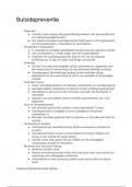
Fetal Head (NORMAL)
8 CRANIAL BONES, SUTURES, & FONTANELLES
____________ connects the 8 cranial bones
sutures
a flexible, connective tissue that lies b/t the cranial bones. (penny ch 24 key term)
Suture (skull):
Fetal sutures SONO (1)
hypoechoic spaces b/t cranial bones.
Name the 8 cranial Bones
Frontal bone
Parietal bones (2)
Temporal bones (2)
Occipital bone
Sphenoid bone
Ethmoid bone
soft spots aka __________
fontanelles
fontanelles are aka ___________
soft spots
__________ are utilized as sonographic windows for neonatal head ultrasound exams
Fontanelles
____________ ____________ is the opening in the base of the cranium through which
the spinal cord travels
Foramen magnum
INTRO
By week ______ the neural plate, the structure that will form the central nervous
system, has developed.
- The neural plate will give rise to the _____________ which will become the spine and
brain.
4.5 weeks
neural tube,
Initially, the brain is divided into 3 primary vesicles. These vesicles continue to develop
and form critical brain structures. What are the 3 primary vesicles?
Prosencephalon (forebrain)
Mesencephalon (midbrain)
Rhombencephalon (hindbrain)
Prosencephalon is aka _______
forebrain
Mesencephalon is aka _________
midbrain
Rhombencephalon is aka __________.
hindbrain
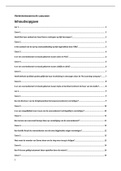
axial view of brain (aka transverse plane)
**axial view is labeled horizontal view in the image**
,Coronal view of brain
What are the 3 axial views of the fetal head?
List in order of most superior to inferior*
Transventricular (aka Ventricular) view (most superior)
Transthalamic (aka thalamic) view
Transcerebellar (aka Cerebellar) view (most inferior)
Which of the following axial views is the BPD/HC/OFD/CI measurements taken?
a. Transventricular
b. Transthalamic
c. Transcerebellar
d. Sagittal
b. Transthalamic (axial)
Which of the following axial views is the LV measurement taken?
a. Transventricular
b. Transthalamic
c. Transcerebellar
d. Sagittal
a. Transventricular (axial)
Which of the following axial views is the Cerebellar length (aka TCD), Cisterna Magna,
& Nuchal fold thickness measurements taken?
a. Transventricular
b. Transthalamic
c. Transcerebellar
d. Sagittal
c. Transcerebellar (Axial)
What intracranial structures will be see in a transventricular view of the fetal head?
LVs (Frontal horns, Occipital horns, body, atrium, etc)
Choroid plexus
Falx cerebri
What intracranial structures will be see in a transthalamic view of the fetal head?
Falx cerebri
CSP
Thalamus / 3rd Ventricle
What anatomic structures will be see in a transcerebellar view of the fetal head?
Cerebellum
Cisterna Magna
Nuchal Fold
(Occipital bone)
, What divides the cerebrum into right & left hemispheres?
falx cerebri (VCU)
interhemispheric fissure (penny)
Both answers are correct bc the falx cerebri which lies within the interhemispheric
fissure
What is the Largest part of brain?
cerebrum
Cerebrum contains multiple ________ & ________.
sulci & gyri
folds in the cerebral cortex. (penny ch 24 key term)
Gyri
grooves within the brain (penny ch 24 key term)
Sulci
sulci and gyri can be found in what structure?
cerebrum
Cerebrum is covered by 3 layers of meninges. What are they?
Pia mater (innermost layer of meninges)
Arachnoid membrane (middle layer of the meninges)
Dura Mater (dense, outermost layer)
What is the innermost layer of the meninges?
Pia Mater
What is the middle layer of meninges?
Arachnoid membrane
What is the dense, outer layer of meninges?
Dura Mater
__________: 3 protective tissue layers that cover the brain and the spinal cord. (p.583
Meninges
double fold of dura mater that separates the cerebral hemispheres.
Falx Cerebri (aka cerebral falx):
Falx cerebri is aka __________
cerebral falx
Located w/in interhemispheric fissure (p.583)
Falx cerebri
groove within the midline of the brain that divides the two cerebral hemispheres. (penny
ch 24 key term)
Interhemispheric Fissure:
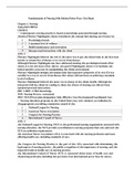
What anatomic structure?
- In the axial plane, seen as hyperechoic line in midline skull
Falx cerebri
Visualization of falx implies...
separation of the cerebrum (aka cerebral hemispheres)
8 CRANIAL BONES, SUTURES, & FONTANELLES
____________ connects the 8 cranial bones
sutures
a flexible, connective tissue that lies b/t the cranial bones. (penny ch 24 key term)
Suture (skull):
Fetal sutures SONO (1)
hypoechoic spaces b/t cranial bones.
Name the 8 cranial Bones
Frontal bone
Parietal bones (2)
Temporal bones (2)
Occipital bone
Sphenoid bone
Ethmoid bone
soft spots aka __________
fontanelles
fontanelles are aka ___________
soft spots
__________ are utilized as sonographic windows for neonatal head ultrasound exams
Fontanelles
____________ ____________ is the opening in the base of the cranium through which
the spinal cord travels
Foramen magnum
INTRO
By week ______ the neural plate, the structure that will form the central nervous
system, has developed.
- The neural plate will give rise to the _____________ which will become the spine and
brain.
4.5 weeks
neural tube,
Initially, the brain is divided into 3 primary vesicles. These vesicles continue to develop
and form critical brain structures. What are the 3 primary vesicles?
Prosencephalon (forebrain)
Mesencephalon (midbrain)
Rhombencephalon (hindbrain)
Prosencephalon is aka _______
forebrain
Mesencephalon is aka _________
midbrain
Rhombencephalon is aka __________.
hindbrain
axial view of brain (aka transverse plane)
**axial view is labeled horizontal view in the image**
,Coronal view of brain
What are the 3 axial views of the fetal head?
List in order of most superior to inferior*
Transventricular (aka Ventricular) view (most superior)
Transthalamic (aka thalamic) view
Transcerebellar (aka Cerebellar) view (most inferior)
Which of the following axial views is the BPD/HC/OFD/CI measurements taken?
a. Transventricular
b. Transthalamic
c. Transcerebellar
d. Sagittal
b. Transthalamic (axial)
Which of the following axial views is the LV measurement taken?
a. Transventricular
b. Transthalamic
c. Transcerebellar
d. Sagittal
a. Transventricular (axial)
Which of the following axial views is the Cerebellar length (aka TCD), Cisterna Magna,
& Nuchal fold thickness measurements taken?
a. Transventricular
b. Transthalamic
c. Transcerebellar
d. Sagittal
c. Transcerebellar (Axial)
What intracranial structures will be see in a transventricular view of the fetal head?
LVs (Frontal horns, Occipital horns, body, atrium, etc)
Choroid plexus
Falx cerebri
What intracranial structures will be see in a transthalamic view of the fetal head?
Falx cerebri
CSP
Thalamus / 3rd Ventricle
What anatomic structures will be see in a transcerebellar view of the fetal head?
Cerebellum
Cisterna Magna
Nuchal Fold
(Occipital bone)
, What divides the cerebrum into right & left hemispheres?
falx cerebri (VCU)
interhemispheric fissure (penny)
Both answers are correct bc the falx cerebri which lies within the interhemispheric
fissure
What is the Largest part of brain?
cerebrum
Cerebrum contains multiple ________ & ________.
sulci & gyri
folds in the cerebral cortex. (penny ch 24 key term)
Gyri
grooves within the brain (penny ch 24 key term)
Sulci
sulci and gyri can be found in what structure?
cerebrum
Cerebrum is covered by 3 layers of meninges. What are they?
Pia mater (innermost layer of meninges)
Arachnoid membrane (middle layer of the meninges)
Dura Mater (dense, outermost layer)
What is the innermost layer of the meninges?
Pia Mater
What is the middle layer of meninges?
Arachnoid membrane
What is the dense, outer layer of meninges?
Dura Mater
__________: 3 protective tissue layers that cover the brain and the spinal cord. (p.583
Meninges
double fold of dura mater that separates the cerebral hemispheres.
Falx Cerebri (aka cerebral falx):
Falx cerebri is aka __________
cerebral falx
Located w/in interhemispheric fissure (p.583)
Falx cerebri
groove within the midline of the brain that divides the two cerebral hemispheres. (penny
ch 24 key term)
Interhemispheric Fissure:
What anatomic structure?
- In the axial plane, seen as hyperechoic line in midline skull
Falx cerebri
Visualization of falx implies...
separation of the cerebrum (aka cerebral hemispheres)