ADVANCED PROTEIN
TECHNOLOGY AND
PROTEOME ANALYSIS
1st Master Biomedical Sciences
Moleculaire mechanismen van ziekten
Gedoceerd door: X. Van Ostade en K. Boonen
UAntwerpen
,H1: Post translational modifications (PTMs)
PTM’s are very important à proteomics can be used to detect PTM’s on proteins!
1. CYSTINE BRIDGE FORMATION (= FIRST PTM)
Cys$ne bridges (= disulfide bonds): common PTM
• Crucial role in stabiliza@on of protein structures à vital for maintaining conforma@ons and func@on
• Bonds form between the sulfur (-S) atoms of two cysteine residues within a protein
Difference between (–SH)₂ and –S–S–
• (–SH)₂: Refers to the presence of two thiol (-SH) groups, one on each cysteine residue.
• –S–S–: Represents the disulfide bond (cys@ne), where the two thiol groups from different cysteine residues
have undergone an oxida@on reac@on to form a covalent linkage.
• The mass difference between free cysteine (–SH) and cys@ne (–S–S–) is 2 Da.
o This difference between free thiol groups and disulfide bonds is small, making it challenging to detect
without high-resolu$on mass spectrometry.
Deriva$za$on of Free SulEydryl Groups:
• Reduc$on of the protein with agents like β-mercaptoethanol breaks disulfide bonds, resul@ng in free
sulPydryl groups.
• Before and aQer reduc@on, free sulPydryl groups (–SH) can be chemically modified.
• Para-hydroxy mercury benzoate (pHMB) is one such chemical reagent used for deriva@za@on
o Adding a mass shiQ of 321 Da.
• Deriva$za$on with pHMB or similar reagents before and aQer reduc@on allows for the iden@fica@on of
cysteines involved in disulfide bridge forma@on.
AQer deriva@za@on and trypsin diges@on (to break the protein into smaller pep@des), mass spectrometry sequencing
can reveal which cysteines were originally involved in forming the disulfide bonds.
• The modifica@on and resul@ng mass shiQ (e.g., 321 Da from pHMB deriva@za@on) enable the specific
detec@on of cysteine residues par@cipa@ng in disulfide bonding.
• The exact posi@ons of the cysteines can be determined by analyzing the mass shiQ paWern in the mass
spectrometry data, correla@ng it with the known pep@de sequences.
2. PHOSPHOPROTEOMICS (= SECOND PTM)
2.1. GENERAL
Phosphoproteomics
• Subfield of proteomics
• Focused on the iden@fica@on, quan@fica@on, and analysis of phosphorylated proteins and their
phosphoryla@on sites.
• Plays a vital role in understanding signaling pathways and cellular regula@on because phosphoryla@on is a
key post-transla@onal modifica@on (PTM) involved in many biological processes.
1
,Prevalence and complexity
• High occurrence: It is es@mated that > 50% of all proteins are phosphorylated at least once during their
life@me, with >100,000 phosphoryla@on sites across the proteome.
• Combinatorial complexity: Many proteins can have mul@ple phosphoryla@on sites, leading to diverse
phosphoryla@on combina@ons (e.g., singly, doubly, or mul@ply phosphorylated states).
Key challenges
• Low abundance and stoichiometry of phosphorylated proteins
o OQen only a small frac@on of the protein is phosphorylated à problem with sensi@vity (detec@on)
• Dynamic regula@on of phosphoryla@on
o Phosphoryla@on is reversible and @ghtly regulated by kinases/phosphatases
o Phosphoryla@on paWerns can change rapidly in response to s@muli
• Ioniza@on suppression
o Nega@ve charge of phosphate groups can reduce ioniza@on efficiency of MS
o This leads to low signal intensi@es
Workflow
1. Enrichment: Techniques to isolate phosphorylated pep@des.
2. Mass Spectrometry: Advanced methods (e.g., ETD, CID) iden@fy and localize phosphoryla@on sites.
3. Data Analysis: Determines phosphoryla@on paWerns and links them to biological pathways.
2.2. ENRICHMENT OF PHOSPHOPEPTIDES
• Essen@al in phosphoproteomics: phosphorylated proteins and pep@des are present at low abundance and
oQen at low stoichiometry compared to non-phosphorylated forms.
• Without enrichment, the signals from phosphorylated pep@des are typically overshadowed by the more
abundant non-phosphorylated pep@des during mass spectrometry (MS) analysis.
2.2.1. IMMUNO AFFINITY CHROMATOGRAPHY (IAC)
Immuno Affinity Chromatography (IAC)
• Technique to selec@vely capture and isolate specific proteins or pep@des based on their an@genic proper@es
• Using an@bodies (Ab)
An$bodies to capture phosphorylated pep@des
• Ab against pY (phosphotyrosine), pS (phosphoserine), and pT (phosphothreonine) are commonly used
• pS and pT an$bodies: less specific due to poten@al cross-reac@vity with other acidic AA (like Asp and Glu).
• Ab against kinase consensus sequences can also be used for broader specificity.
Applica$ons:
• OQen used in combina$on with other methods like IMAC (immobilized metal affinity chromatography) or
TiO₂ (@tanium dioxide chromatography) to enhance specificity and capture phosphorylated pep@des more
efficiently.
2
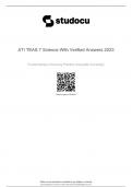
, 2.2.2. IMMOBILIZED METAL AFFINITY CHROMATOGRAPHY (IMAC)
This technique takes advantage of the affinity between nega@vely charged phosphate groups on pep@des and metal
ions immobilized on a sta@onary phase.
Prepara$on of the Sta$onary Phase:
• Metal Chela@on: Metal ions (e.g., Fe³⁺, Ga³⁺) are chelated (chemically bound) to nitrilotriace@c acid (NTA)
ligands, which are pre-aWached to beads.
• This creates a stable metal-NTA complex that forms the sta@onary phase for chromatography.
Binding of Phosphopep$des:
• Phosphopep@des have (-) charged phosphate groups that interact strongly with the (+) charged metal ions.
• The binding is driven by the electrosta@c interac@on between the phosphate groups and the metal centers.
• Non-specific Binding: Other acidic pep@des (rich in aspar@c or glutamic acid residues) or non-phosphorylated
pep@des with anionic features may also bind to some extent, reducing specificity.
Preference for Mul$phosphorylated Pep$des:
• Pep@des with mul@ple phosphate groups exhibit stronger binding due to their higher nega@ve charge and
greater affinity for the sta@onary phase.
Elu$on of Bound Pep$des:
• Elu@on is achieved under basic condi$ons (high pH), which disrupts the interac@on between the phosphate
groups and the metal ions.
2.2.3. VARIATION: SEQUENCTIAL IMAC (SMAC)
3
TECHNOLOGY AND
PROTEOME ANALYSIS
1st Master Biomedical Sciences
Moleculaire mechanismen van ziekten
Gedoceerd door: X. Van Ostade en K. Boonen
UAntwerpen
,H1: Post translational modifications (PTMs)
PTM’s are very important à proteomics can be used to detect PTM’s on proteins!
1. CYSTINE BRIDGE FORMATION (= FIRST PTM)
Cys$ne bridges (= disulfide bonds): common PTM
• Crucial role in stabiliza@on of protein structures à vital for maintaining conforma@ons and func@on
• Bonds form between the sulfur (-S) atoms of two cysteine residues within a protein
Difference between (–SH)₂ and –S–S–
• (–SH)₂: Refers to the presence of two thiol (-SH) groups, one on each cysteine residue.
• –S–S–: Represents the disulfide bond (cys@ne), where the two thiol groups from different cysteine residues
have undergone an oxida@on reac@on to form a covalent linkage.
• The mass difference between free cysteine (–SH) and cys@ne (–S–S–) is 2 Da.
o This difference between free thiol groups and disulfide bonds is small, making it challenging to detect
without high-resolu$on mass spectrometry.
Deriva$za$on of Free SulEydryl Groups:
• Reduc$on of the protein with agents like β-mercaptoethanol breaks disulfide bonds, resul@ng in free
sulPydryl groups.
• Before and aQer reduc@on, free sulPydryl groups (–SH) can be chemically modified.
• Para-hydroxy mercury benzoate (pHMB) is one such chemical reagent used for deriva@za@on
o Adding a mass shiQ of 321 Da.
• Deriva$za$on with pHMB or similar reagents before and aQer reduc@on allows for the iden@fica@on of
cysteines involved in disulfide bridge forma@on.
AQer deriva@za@on and trypsin diges@on (to break the protein into smaller pep@des), mass spectrometry sequencing
can reveal which cysteines were originally involved in forming the disulfide bonds.
• The modifica@on and resul@ng mass shiQ (e.g., 321 Da from pHMB deriva@za@on) enable the specific
detec@on of cysteine residues par@cipa@ng in disulfide bonding.
• The exact posi@ons of the cysteines can be determined by analyzing the mass shiQ paWern in the mass
spectrometry data, correla@ng it with the known pep@de sequences.
2. PHOSPHOPROTEOMICS (= SECOND PTM)
2.1. GENERAL
Phosphoproteomics
• Subfield of proteomics
• Focused on the iden@fica@on, quan@fica@on, and analysis of phosphorylated proteins and their
phosphoryla@on sites.
• Plays a vital role in understanding signaling pathways and cellular regula@on because phosphoryla@on is a
key post-transla@onal modifica@on (PTM) involved in many biological processes.
1
,Prevalence and complexity
• High occurrence: It is es@mated that > 50% of all proteins are phosphorylated at least once during their
life@me, with >100,000 phosphoryla@on sites across the proteome.
• Combinatorial complexity: Many proteins can have mul@ple phosphoryla@on sites, leading to diverse
phosphoryla@on combina@ons (e.g., singly, doubly, or mul@ply phosphorylated states).
Key challenges
• Low abundance and stoichiometry of phosphorylated proteins
o OQen only a small frac@on of the protein is phosphorylated à problem with sensi@vity (detec@on)
• Dynamic regula@on of phosphoryla@on
o Phosphoryla@on is reversible and @ghtly regulated by kinases/phosphatases
o Phosphoryla@on paWerns can change rapidly in response to s@muli
• Ioniza@on suppression
o Nega@ve charge of phosphate groups can reduce ioniza@on efficiency of MS
o This leads to low signal intensi@es
Workflow
1. Enrichment: Techniques to isolate phosphorylated pep@des.
2. Mass Spectrometry: Advanced methods (e.g., ETD, CID) iden@fy and localize phosphoryla@on sites.
3. Data Analysis: Determines phosphoryla@on paWerns and links them to biological pathways.
2.2. ENRICHMENT OF PHOSPHOPEPTIDES
• Essen@al in phosphoproteomics: phosphorylated proteins and pep@des are present at low abundance and
oQen at low stoichiometry compared to non-phosphorylated forms.
• Without enrichment, the signals from phosphorylated pep@des are typically overshadowed by the more
abundant non-phosphorylated pep@des during mass spectrometry (MS) analysis.
2.2.1. IMMUNO AFFINITY CHROMATOGRAPHY (IAC)
Immuno Affinity Chromatography (IAC)
• Technique to selec@vely capture and isolate specific proteins or pep@des based on their an@genic proper@es
• Using an@bodies (Ab)
An$bodies to capture phosphorylated pep@des
• Ab against pY (phosphotyrosine), pS (phosphoserine), and pT (phosphothreonine) are commonly used
• pS and pT an$bodies: less specific due to poten@al cross-reac@vity with other acidic AA (like Asp and Glu).
• Ab against kinase consensus sequences can also be used for broader specificity.
Applica$ons:
• OQen used in combina$on with other methods like IMAC (immobilized metal affinity chromatography) or
TiO₂ (@tanium dioxide chromatography) to enhance specificity and capture phosphorylated pep@des more
efficiently.
2
, 2.2.2. IMMOBILIZED METAL AFFINITY CHROMATOGRAPHY (IMAC)
This technique takes advantage of the affinity between nega@vely charged phosphate groups on pep@des and metal
ions immobilized on a sta@onary phase.
Prepara$on of the Sta$onary Phase:
• Metal Chela@on: Metal ions (e.g., Fe³⁺, Ga³⁺) are chelated (chemically bound) to nitrilotriace@c acid (NTA)
ligands, which are pre-aWached to beads.
• This creates a stable metal-NTA complex that forms the sta@onary phase for chromatography.
Binding of Phosphopep$des:
• Phosphopep@des have (-) charged phosphate groups that interact strongly with the (+) charged metal ions.
• The binding is driven by the electrosta@c interac@on between the phosphate groups and the metal centers.
• Non-specific Binding: Other acidic pep@des (rich in aspar@c or glutamic acid residues) or non-phosphorylated
pep@des with anionic features may also bind to some extent, reducing specificity.
Preference for Mul$phosphorylated Pep$des:
• Pep@des with mul@ple phosphate groups exhibit stronger binding due to their higher nega@ve charge and
greater affinity for the sta@onary phase.
Elu$on of Bound Pep$des:
• Elu@on is achieved under basic condi$ons (high pH), which disrupts the interac@on between the phosphate
groups and the metal ions.
2.2.3. VARIATION: SEQUENCTIAL IMAC (SMAC)
3