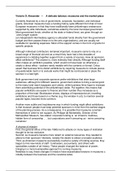
Task 1 – Brain development and structures
Different views of the brain
Directions in nervous system are
described in relation to the
orientation of the spinal cord:
- Anterior-posterior axis
• Anterior/frontal/rostral: in front
or towards the nose end
• Posterior/caudal: toward the
back or the tail
- Dorsal-ventral axis
• Dorsal/superior: toward
surface of back or top of head
• Ventral/inferior: toward
surface of chest or bottom of
head
➔ Superior is used to refer to
top of head, inferior is
used to refer to bottom of
head
- Medial-lateral axis
• Medial: close to center or
toward midline of body
• Lateral: to the side or away
from midline toward body’s lateral surfaces
➔ Dorsal and ventral in animals are anterior and posterior in upright humans, but if humans
stand on all fours, the orientation of spinal cord is similar to that of other animals
- Proximal: closer to CNS
- Distal: farther from CNS
- Horizontal plane (transverse, axial):
divides brain into superior and inferior (dorsal
view)
- Frontal/coronal plane: divides brain in front
and back, parallel to forehead (frontal view)
- Sagittal plane: divides brain in left and
right (medial view)
- Midsagittal plane: divides brain in 2
hemispheres
- Cross section: section cut at long,
narrow structure (spinal cord or nerve)
Pagina 1 van 100
,Nervous system
- Central Nervous System (CNS) = division of nervous system located within skull (brain) and
spine (spinal cord)
- Peripheral Nervous System (PNS) = division of nervous system located outside skull & spine
• Somatic Nervous System (SNS) = interacts with external environment
➢ Afferent nerves: carry sensory signals from the skin, skeletal muscles, eyes, ears, etc.
to CNS
➢ Efferent nerves: carry motor signals from CNS to skeletal muscles
➔ Most of nerves of PNS project from spinal cord, but there are 12 pairs of exception:
➢ Cranial nerves (12) = serve sensory and motor functions of head and neck region –
attached to ventral surface of brain – sensory nerves, optic nerves, sensory and
motor fibers (vagus nerve = regulates functions of organs)
➢ Spinal nerves = begin at junction of dorsal and ventral roots of spinal cord
• Autonomic Nervous System (ANS) = regulates body’s internal environment
➢ Afferent nerves: carry sensory signals from internal organs to CNS
➢ Efferent nerves: carry motor signals from CNS to internal organs
o Sympathetic nerves: autonomic motor nerves that project from CNS in lumbar
(small of the back) and thoracic (chest area) regions of spinal cord
o Parasympathetic nerves: autonomic motor nerves that project from brain and
sacral (lower back) region of spinal cord
▪ Sympathetic nerves stimulate, organize and mobilize energy resources in
threatening situations and parasympathetic nerves act to conserve energy
(increase body’s supply of stored energy and return systems to resting state
following activation by sympathetic nervous system)
▪ Sympathetic changes are indicative of psychological arousal and
parasympathetic changes are indicative of psychological relaxation
▪ Each autonomic target organ receives opposing sympathetic and
parasympathetic input and its activity is thus controlled by relative levels of
sympathetic and parasympathetic activity
Pagina 2 van 100
,Spinal cord
Function: relay sensory information to brain from body, relay motor information from brain to
body and coordinate reflexive behaviors
The spinal cord comprises 2 different areas:
- Inner H-shaped core of gray matter (composed of cell bodies and unmyelinated axons)
- Surrounding area of white matter (composed of myelinated axons)
- Dorsal horns: 2 dorsal arms of spinal gray matter
- Ventral horns: 2 ventral arms of spinal gray matter
- Pairs of spinal nerves are attached to spinal cord (1 on left, 1 on right) at 31 levels of spine –
each of these 62 spinal nerves divide as it nears the cord, and its axons are joined to cord
via dorsal root or ventral root
- Dorsal root axons: sensory (afferent) unipolar
neurons with their cell bodies grouped together just
outside cord to form dorsal root ganglia – many of
their synaptic terminals are in dorsal horns of spinal
gray matter
- Ventral root axons: motor (efferent) multipolar
neurons with their cell bodies in ventral horns – those
that are part of SNS project to skeletal muscles,
those that are part of ANS project to ganglia, where
they synapse on neurons that project to internal
organs
Development of brain
Protective sheaths around the brain and spinal cord are referred to as meninges:
- Dura mater: durable, thick, though and flexible but unstretchable
- Arachnoid membrane: soft and spongy
- Pia mater: smaller surface blood vessels of the brain and spinal cord are contained within
this layer
- Subarachnoid space: between pia mater and arachnoid membrane: filled with CSF
Pagina 3 van 100
, PNS is covered with 2 layers of meninges:
- Middle layer (arachnoid membraine): covers only brain and spinal cord, with its associated
pool of CSF
- Outer and inner layers (dura mater and pia mater): fuse and form sheath that covers spinal
and cranial nerves and peripheral ganglia
Evolution of human brain
- There is no clear relationship between overall human brain size and intelligence
- It is more informative to consider evolution of the brain stem separately from evolution of the
cerebrum (cerebral hemispheres)
- Brain stem = regulates reflex activities critical for survival (heart rate, respiration, blood
glucose level)
- Cerebrum = involved in complex adaptive processes (learning, perception and motivation)
➔ The brain has increased in size during evolution
➔ Most of the increase in size has occurred in cerebrum
➔ An increase in number of convolutions (folds on cerebral cortex) has greatly increased
the surface area of cerebral cortex, the outermost layer of cerebral tissue
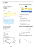
Development of nervous system: from ectoderm plate, to neural tube, to 3 interconnected
chambres
Ectoderm begins to develop at embryonic day 18. As more cells are added to neural plate, the
edges of the plate begin to curl together, forming neural tube at embryonic day 20. The neural
tube fuses together at embryonic day 28. The tube then begins to develop 2 interconnected
chambers that will become the forebrain, midbrain and hindbrain.
Prenatal development
Cerebral cortex of humans develops from the inside out, as progenitor cells from ventricular
system move into their final positions in the cortex. Initial brain development occurs during
period of symmetrical division, followed by period of asymmetrical division when embryo is 7
weeks old. Asymmetrical division last for 3 months and produces radial glia, which help to
establish the cortex. Once neurons have migrated to their final location, they begin forming
connections with other neurons. The axons of 50% of new neurons don’t form synaptic
connections, so they die by apoptosis. Genetic change, personal experience and neurogenesis
influence postnatal brain development.
5 major divisions of the brain
Early development of brain:
- In vertebrate embryo, the tissue that eventually develops into CNS is recognizable as fluid-
filling tube
Pagina 4 van 100
Different views of the brain
Directions in nervous system are
described in relation to the
orientation of the spinal cord:
- Anterior-posterior axis
• Anterior/frontal/rostral: in front
or towards the nose end
• Posterior/caudal: toward the
back or the tail
- Dorsal-ventral axis
• Dorsal/superior: toward
surface of back or top of head
• Ventral/inferior: toward
surface of chest or bottom of
head
➔ Superior is used to refer to
top of head, inferior is
used to refer to bottom of
head
- Medial-lateral axis
• Medial: close to center or
toward midline of body
• Lateral: to the side or away
from midline toward body’s lateral surfaces
➔ Dorsal and ventral in animals are anterior and posterior in upright humans, but if humans
stand on all fours, the orientation of spinal cord is similar to that of other animals
- Proximal: closer to CNS
- Distal: farther from CNS
- Horizontal plane (transverse, axial):
divides brain into superior and inferior (dorsal
view)
- Frontal/coronal plane: divides brain in front
and back, parallel to forehead (frontal view)
- Sagittal plane: divides brain in left and
right (medial view)
- Midsagittal plane: divides brain in 2
hemispheres
- Cross section: section cut at long,
narrow structure (spinal cord or nerve)
Pagina 1 van 100
,Nervous system
- Central Nervous System (CNS) = division of nervous system located within skull (brain) and
spine (spinal cord)
- Peripheral Nervous System (PNS) = division of nervous system located outside skull & spine
• Somatic Nervous System (SNS) = interacts with external environment
➢ Afferent nerves: carry sensory signals from the skin, skeletal muscles, eyes, ears, etc.
to CNS
➢ Efferent nerves: carry motor signals from CNS to skeletal muscles
➔ Most of nerves of PNS project from spinal cord, but there are 12 pairs of exception:
➢ Cranial nerves (12) = serve sensory and motor functions of head and neck region –
attached to ventral surface of brain – sensory nerves, optic nerves, sensory and
motor fibers (vagus nerve = regulates functions of organs)
➢ Spinal nerves = begin at junction of dorsal and ventral roots of spinal cord
• Autonomic Nervous System (ANS) = regulates body’s internal environment
➢ Afferent nerves: carry sensory signals from internal organs to CNS
➢ Efferent nerves: carry motor signals from CNS to internal organs
o Sympathetic nerves: autonomic motor nerves that project from CNS in lumbar
(small of the back) and thoracic (chest area) regions of spinal cord
o Parasympathetic nerves: autonomic motor nerves that project from brain and
sacral (lower back) region of spinal cord
▪ Sympathetic nerves stimulate, organize and mobilize energy resources in
threatening situations and parasympathetic nerves act to conserve energy
(increase body’s supply of stored energy and return systems to resting state
following activation by sympathetic nervous system)
▪ Sympathetic changes are indicative of psychological arousal and
parasympathetic changes are indicative of psychological relaxation
▪ Each autonomic target organ receives opposing sympathetic and
parasympathetic input and its activity is thus controlled by relative levels of
sympathetic and parasympathetic activity
Pagina 2 van 100
,Spinal cord
Function: relay sensory information to brain from body, relay motor information from brain to
body and coordinate reflexive behaviors
The spinal cord comprises 2 different areas:
- Inner H-shaped core of gray matter (composed of cell bodies and unmyelinated axons)
- Surrounding area of white matter (composed of myelinated axons)
- Dorsal horns: 2 dorsal arms of spinal gray matter
- Ventral horns: 2 ventral arms of spinal gray matter
- Pairs of spinal nerves are attached to spinal cord (1 on left, 1 on right) at 31 levels of spine –
each of these 62 spinal nerves divide as it nears the cord, and its axons are joined to cord
via dorsal root or ventral root
- Dorsal root axons: sensory (afferent) unipolar
neurons with their cell bodies grouped together just
outside cord to form dorsal root ganglia – many of
their synaptic terminals are in dorsal horns of spinal
gray matter
- Ventral root axons: motor (efferent) multipolar
neurons with their cell bodies in ventral horns – those
that are part of SNS project to skeletal muscles,
those that are part of ANS project to ganglia, where
they synapse on neurons that project to internal
organs
Development of brain
Protective sheaths around the brain and spinal cord are referred to as meninges:
- Dura mater: durable, thick, though and flexible but unstretchable
- Arachnoid membrane: soft and spongy
- Pia mater: smaller surface blood vessels of the brain and spinal cord are contained within
this layer
- Subarachnoid space: between pia mater and arachnoid membrane: filled with CSF
Pagina 3 van 100
, PNS is covered with 2 layers of meninges:
- Middle layer (arachnoid membraine): covers only brain and spinal cord, with its associated
pool of CSF
- Outer and inner layers (dura mater and pia mater): fuse and form sheath that covers spinal
and cranial nerves and peripheral ganglia
Evolution of human brain
- There is no clear relationship between overall human brain size and intelligence
- It is more informative to consider evolution of the brain stem separately from evolution of the
cerebrum (cerebral hemispheres)
- Brain stem = regulates reflex activities critical for survival (heart rate, respiration, blood
glucose level)
- Cerebrum = involved in complex adaptive processes (learning, perception and motivation)
➔ The brain has increased in size during evolution
➔ Most of the increase in size has occurred in cerebrum
➔ An increase in number of convolutions (folds on cerebral cortex) has greatly increased
the surface area of cerebral cortex, the outermost layer of cerebral tissue
Development of nervous system: from ectoderm plate, to neural tube, to 3 interconnected
chambres
Ectoderm begins to develop at embryonic day 18. As more cells are added to neural plate, the
edges of the plate begin to curl together, forming neural tube at embryonic day 20. The neural
tube fuses together at embryonic day 28. The tube then begins to develop 2 interconnected
chambers that will become the forebrain, midbrain and hindbrain.
Prenatal development
Cerebral cortex of humans develops from the inside out, as progenitor cells from ventricular
system move into their final positions in the cortex. Initial brain development occurs during
period of symmetrical division, followed by period of asymmetrical division when embryo is 7
weeks old. Asymmetrical division last for 3 months and produces radial glia, which help to
establish the cortex. Once neurons have migrated to their final location, they begin forming
connections with other neurons. The axons of 50% of new neurons don’t form synaptic
connections, so they die by apoptosis. Genetic change, personal experience and neurogenesis
influence postnatal brain development.
5 major divisions of the brain
Early development of brain:
- In vertebrate embryo, the tissue that eventually develops into CNS is recognizable as fluid-
filling tube
Pagina 4 van 100