Week 2:
Respiratory Disorders and Alterations in Acid/Base Balance,
Fluid and Electrolytes
Chapter 35: Structure and Function of the Pulmonary System
Tara Morgan
• The primary function of the pulmonary system is the exchange of gases between
the environmental air and the blood.
• There are three steps in this process:
○ Ventilation, the movement of air into and out of the lungs
○ Diffusion, the movement of gases between air spaces in the lungs and the
bloodstream
○ Perfusion, the movement of blood into and out of the capillary beds of the
lungs to body organs and tissues
• The first two functions (ventilation and diffusion) are carried out by the
pulmonary system and the third (perfusion) by the cardiovascular system.
• Normally the pulmonary system functions efficiently under a variety of conditions
and with little energy expenditure.
Structures of the Pulmonary System
• The pulmonary system includes two lungs and the upper and lower airways, and
the blood vessels that serve them; the chest wall, or thoracic cage; and the
diaphragm.
• The lungs are divided into lobes:
○ 3 in the right lung (upper, middle, lower)
○ 2 in the left lung (upper, lower)
• Each lobe is further divided into segments and lobules.
• The mediastinum is the space between the lungs and contains the heart, great
vessels, and esophagus.
• A set of conducting airways, or bronchi, delivers air to each section of the lung.
• The diaphragm is a dome-shaped muscle that separates the thoracic and
abdominal cavities and is involved in ventilation.
•
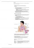
• FIGURE 35.1 Structural Plan of the Respiratory System. The inset shows alveolar
sacs where the interchange of oxygen and carbon dioxide takes place through the
walls of the grapelike alveoli. Capillaries surround the alveoli.
• The lungs are protected from a variety of exogenous contaminants by a series of
mechanical and cellular defenses.
• These defense mechanisms are so effective that in the healthy individual,
contamination of the lung tissue itself, particularly by infectious agents, is rare.
STRUCTURE MECHANISM OF DEFENSE
OR
SUBSTANCE
Upper Maintains constant temperature and humidification of gas entering
respiratory tract the lungs; traps and removes foreign particles, some bacteria, and
mucosa noxious gases from inspired air
Nasal hairs and Trap and remove foreign particles, some bacteria, and noxious
turbinates gases from inspired air
, Branching Disrupt laminar flow and enhance deposition of particles and
airways pathogens on ciliated mucosa
Mucous blanket Protects trachea and bronchi from injury; traps most foreign
particles and bacteria that reach the lower airways
Innate immune Lysozyme, lactoferrin, defensins, collectins (surfactant protein A
proteins [SP-A] and surfactant protein D [SP-D]), and immunoglobulin A
(IgA); recognize and promote killing of pathogens
Cilia Propel mucous blanket and entrapped particles toward the
oropharynx, where they can be swallowed or expectorated
Alveolar Ingest and remove bacteria and other foreign material from alveoli
macrophages by phagocytosis (see Chapter 7)
Surfactant Enhances phagocytosis of pathogens and allergens in alveoli;
down-regulates inflammatory responses
Irritant receptors Stimulation by chemical or mechanical irritants triggers sneeze
in nares reflex, which results in rapid removal of irritants from nasal
(nostrils) passages
Irritant receptors Stimulation by chemical or mechanical irritants triggers cough
in trachea and reflex, which results in removal of irritants from the trachea and
large airways large airways
Conducting Airways
• The conducting airways provide a passage for the movement of air into and out of the
gas-exchange structures of the lung.
• The nasopharynx, oropharynx, and related structures are called the upper airway.
• Lined with ciliated mucosa with a rich vascular supply that warms and humidifies
inspired air and removes foreign particles from it as it passes into the lungs.
• The mouth and oropharynx provide for ventilation when the nose is obstructed or
when increased flow is required, for example, during exercise. Filtering and
humidifying are not as efficient with mouth breathing.
FIGURE 35.2 Structures of the Upper Airway.
• The larynx connects the upper and lower airways and consists of the endolarynx and
its surrounding triangular-shaped bony and cartilaginous structures.
• The endolarynx is formed by two pairs of folds: the false vocal cords (supraglottis)
and the true vocal cords.
• The slit-shaped space between the true cords forms the glottis.
• The vestibule is the space above the false vocal cords.
• The laryngeal box is formed by three large cartilages
, • Epiglottis
• Thyroid
• Cricoid
○ And three smaller cartilages-connected by ligaments
Arytenoid
Corniculate
Cuneiform
• The supporting cartilages prevent collapse of the larynx during inspiration and
swallowing.
• The internal laryngeal muscles control vocal cord length and tension, and the external
laryngeal muscles move the larynx as a whole.
• The internal muscles contract during swallowing to prevent aspiration into the trachea
and contribute to voice pitch.
• The trachea, which is supported by U-shaped cartilage, connects the larynx to the
bronchi, the conducting airways of the lungs.
• The trachea divides into the two main airways, or bronchi, at the carina
• This area is very sensitive and when stimulated can cause coughing and airway
narrowing.
• The left mainstem bronchus branches from the trachea at about a 45 -degree angle.
The right mainstem bronchus is slightly larger and more vertical than the left
(branches at about a 20- to 30-degree angle from the trachea).
• Aspirated fluids or foreign particles thus tend to enter the right lung rather than
the left.
• The right and left main bronchi enter the lungs at the hila, or ―roots‖ of the lungs,
along with the pulmonary blood and lymphatic vessels.
• From the hila the main bronchi branch into lobar bronchi and then to segmental and
subsegmental bronchi, and finally end at the sixteenth division in the smallest of the
conducting airways, the terminal bronchioles
FIGURE 35.3 Conducting Airways and Respiratory Unit. A, Structures of respiratory
airways. B, Changes in bronchial wall with progressive branching. C, Electron
micrograph of alveoli: long white arrow identifies type II alveolar cells (pneumocytes -
secretes surfactant); short white arrowhead identifies pores of Kohn; red arrow
identifies alveolar capillary. D, Plastic cast of pulmonary capillaries at high
magnification.
• The bronchial walls have 3 layers:
• Epithelial lining
• Smooth muscle layer
• Connective tissue layer
• In the large bronchi (up to approximately the tenth division), the connective tissue
layer contains cartilage.
• The epithelial lining of the bronchi contains single -celled exocrine glands—the mucus-
secreting goblet cells—and ciliated cells.
• High columnar pseudostratified epithelium lines the larger airways and becomes
progressively thinner, changing to columnar cuboidal epithelium in the bronchioles
and squamous epithelium in the alveoli
• The submucosal glands of the bronchial lining produce a mucous blanket that
protects the bronchial epithelium.
• The ciliated epithelial cells rhythmically beat this mucous blanket toward the trachea
, and VVpharynx, VVwhere VVit VVcan VVbe VVswallowed VVor VVexpectorated VVby VVcoughing.
• Toward VVthe VVterminal VVbronchioles, VVciliated VVcells VVand VVgoblet VVcells
VVbecome VVmore VVsparse, V V and VVsmooth VVmuscle VVand VVconnective VVtissue
VVlayers VVthin
Gas-Exchange VVAirways
• The VVconducting VVairways VVterminate VVin VVthe VVrespiratory VV(terminal)
VVbronchioles, VValveolar V V ducts, VVand VValveoli
• These VVthin-walled VVstructures VVparticipate VVin VVgas VVexchange, VVand
VVthe VVclusters VVof V V alveoli VVare VVsometimes VVcalled VVthe VVacinus
• The VVbronchioles VVfrom VVthe VVsixteenth VVthrough VVthe VVtwenty-third
VVdivisions VVcontain V V increasing VVnumbers VVof VValveoli VVand VVare
VVcalled VVrespiratory VVbronchioles.
• The VVwalls VVof VVthe VVrespiratory VVbronchioles VVare VVvery VVthin, VVconsisting
VVof VVan VVepithelial V V layer VVdevoid VVof VVcilia VVand VVgoblet VVcells, VVvery
VVlittle VVsmooth VVmuscle VVfiber, VVand VVa VVvery V V thin VVand VVelastic
VVconnective VVtissue VVlayer.
• These VVbronchioles VVend VVin VValveolar VVducts, VVwhich VVlead VVto
VValveolar VVsacs VVmade VVup V V of VVnumerous VValveoli.
• The VValveoli VVare VVthe VVprimary VVgas-exchange VVunits VVof VVthe VVlung, VVwhere
VVoxygen VVenters VVthe V V blood VVand VVCO2 VVis VVremoved
• Tiny VVpassages VVcalled VVpores VVof VVKohn VVpermit VVsome VVair VVto VVpass
VVthrough VVthe VVsepta VVfrom V V alveolus VVto VValveolus, VVpromoting
VVcollateral VVventilation VVand VVeven VVdistribution VVof VVair V V among VVthe
VValveoli. VVThe VVlungs VVcontain VVapproximately VV50 VVmillion VValveoli VVat
VVbirth VVand V V about VV480 VVmillion VVby VVadulthood.
FIGURE VV35.4 VVAlveoli. VVBronchioles VVsubdivide VVto VVform VVtiny VVtubes VVcalled VValveolar
VVducts VVthat VVend VVin V V clusters VVof VValveoli VVcalled VValveolar VVsacs.
• Lung VVepithelial VVcells VVprovide VVa VVprotective VVinterface VVwith VVthe VVenvironment
• Essential VVfor VVadequate VVgas VVexchange
• Preventing VVentry VVof VVforeign VVagents
• Regulates VVion VVand VVwater VVtransport
• Maintains VVmechanical VVstability VVof VVthe VValveoli
• The VValveolar VVsepta VVconsist VVof VVan VVepithelial VVlayer VVand VVa VVthin,
VVelastic VVbasement V V membrane VVbut VVno VVmuscle VVlayer
• Two VVmajor VVtypes VVof VVepithelial VVcells VV(pneumocytes) VVappear VVin VVthe VValveolus.
• Type VVI VValveolar VVcells VVprovide VVstructure
• Type VVII VValveolar VVcells VVsecrete VVsurfactant, VVa VVlipoprotein VVthat
VVcoats VVthe VVinner V V surface VVof VVthe VValveolus VVand VVfacilitates VVits
VVexpansion VVduring VVinspiration, VVwhich V V lowers VValveolar VVsurface
VVtension VVat VVend-expiration, VVthereby VVpreventing VVlung V V collapse
VV(atelectasis).
• Surfactant
• Contribute VVto VVcontrol VVof VVlung VVinflammation VVby
VVdecreasing VVrelease VVof V V proinflammatory VVmediators
• Prevents VVoxidative VV injury
• Regulates VVthe VVrole VVof VVfibroblasts VVin VVairway VVremodeling.
• Bacteriostatic VVand VVfunction VVas VVopsonins VVin VVpresenting
VVpathogens VVto VValveolar V V macrophages.
• Macrophages VVare VVthe VVmost VVnumerous VVimmune VVcells VVpresent VVin VVthe
VVlung VVenvironment V V and VVprovide VVinnate VVimmune VVdefense VVof VVthe
VVairway VVfrom VVthe VVbronchi VVto VVthe VValveoli.
• In VVthe VValveoli, VValveolar VVmacrophages VVprovide VVprotection VVby VVclearing
VVsurfactant VVfrom VVthe V V lung VVand VVingesting VVforeign VVmaterial VVand
VVpathogens VVthat VVreach VVthe VValveolus, VVpreparing V V these VVsubstances
VVfor VVremoval VVthrough VVthe VVlymphatics VV-Phagocytosis
• Surfactant VVand VValveolar VVmacrophages VVwork VVtogether VVwith VVthe
VVnormal VVpulmonary V V microbiota VVto VVprevent VVlower VVlung VVinfection.
Pulmonary VVand VVBronchial VV Circulation
• The VVpulmonary VVcirculation;
• provides VVan VVextensive VVsurface VVarea VVfor VVgas VVexchange
• delivers VVnutrients VVto VVlung VVtissues
Respiratory Disorders and Alterations in Acid/Base Balance,
Fluid and Electrolytes
Chapter 35: Structure and Function of the Pulmonary System
Tara Morgan
• The primary function of the pulmonary system is the exchange of gases between
the environmental air and the blood.
• There are three steps in this process:
○ Ventilation, the movement of air into and out of the lungs
○ Diffusion, the movement of gases between air spaces in the lungs and the
bloodstream
○ Perfusion, the movement of blood into and out of the capillary beds of the
lungs to body organs and tissues
• The first two functions (ventilation and diffusion) are carried out by the
pulmonary system and the third (perfusion) by the cardiovascular system.
• Normally the pulmonary system functions efficiently under a variety of conditions
and with little energy expenditure.
Structures of the Pulmonary System
• The pulmonary system includes two lungs and the upper and lower airways, and
the blood vessels that serve them; the chest wall, or thoracic cage; and the
diaphragm.
• The lungs are divided into lobes:
○ 3 in the right lung (upper, middle, lower)
○ 2 in the left lung (upper, lower)
• Each lobe is further divided into segments and lobules.
• The mediastinum is the space between the lungs and contains the heart, great
vessels, and esophagus.
• A set of conducting airways, or bronchi, delivers air to each section of the lung.
• The diaphragm is a dome-shaped muscle that separates the thoracic and
abdominal cavities and is involved in ventilation.
•
• FIGURE 35.1 Structural Plan of the Respiratory System. The inset shows alveolar
sacs where the interchange of oxygen and carbon dioxide takes place through the
walls of the grapelike alveoli. Capillaries surround the alveoli.
• The lungs are protected from a variety of exogenous contaminants by a series of
mechanical and cellular defenses.
• These defense mechanisms are so effective that in the healthy individual,
contamination of the lung tissue itself, particularly by infectious agents, is rare.
STRUCTURE MECHANISM OF DEFENSE
OR
SUBSTANCE
Upper Maintains constant temperature and humidification of gas entering
respiratory tract the lungs; traps and removes foreign particles, some bacteria, and
mucosa noxious gases from inspired air
Nasal hairs and Trap and remove foreign particles, some bacteria, and noxious
turbinates gases from inspired air
, Branching Disrupt laminar flow and enhance deposition of particles and
airways pathogens on ciliated mucosa
Mucous blanket Protects trachea and bronchi from injury; traps most foreign
particles and bacteria that reach the lower airways
Innate immune Lysozyme, lactoferrin, defensins, collectins (surfactant protein A
proteins [SP-A] and surfactant protein D [SP-D]), and immunoglobulin A
(IgA); recognize and promote killing of pathogens
Cilia Propel mucous blanket and entrapped particles toward the
oropharynx, where they can be swallowed or expectorated
Alveolar Ingest and remove bacteria and other foreign material from alveoli
macrophages by phagocytosis (see Chapter 7)
Surfactant Enhances phagocytosis of pathogens and allergens in alveoli;
down-regulates inflammatory responses
Irritant receptors Stimulation by chemical or mechanical irritants triggers sneeze
in nares reflex, which results in rapid removal of irritants from nasal
(nostrils) passages
Irritant receptors Stimulation by chemical or mechanical irritants triggers cough
in trachea and reflex, which results in removal of irritants from the trachea and
large airways large airways
Conducting Airways
• The conducting airways provide a passage for the movement of air into and out of the
gas-exchange structures of the lung.
• The nasopharynx, oropharynx, and related structures are called the upper airway.
• Lined with ciliated mucosa with a rich vascular supply that warms and humidifies
inspired air and removes foreign particles from it as it passes into the lungs.
• The mouth and oropharynx provide for ventilation when the nose is obstructed or
when increased flow is required, for example, during exercise. Filtering and
humidifying are not as efficient with mouth breathing.
FIGURE 35.2 Structures of the Upper Airway.
• The larynx connects the upper and lower airways and consists of the endolarynx and
its surrounding triangular-shaped bony and cartilaginous structures.
• The endolarynx is formed by two pairs of folds: the false vocal cords (supraglottis)
and the true vocal cords.
• The slit-shaped space between the true cords forms the glottis.
• The vestibule is the space above the false vocal cords.
• The laryngeal box is formed by three large cartilages
, • Epiglottis
• Thyroid
• Cricoid
○ And three smaller cartilages-connected by ligaments
Arytenoid
Corniculate
Cuneiform
• The supporting cartilages prevent collapse of the larynx during inspiration and
swallowing.
• The internal laryngeal muscles control vocal cord length and tension, and the external
laryngeal muscles move the larynx as a whole.
• The internal muscles contract during swallowing to prevent aspiration into the trachea
and contribute to voice pitch.
• The trachea, which is supported by U-shaped cartilage, connects the larynx to the
bronchi, the conducting airways of the lungs.
• The trachea divides into the two main airways, or bronchi, at the carina
• This area is very sensitive and when stimulated can cause coughing and airway
narrowing.
• The left mainstem bronchus branches from the trachea at about a 45 -degree angle.
The right mainstem bronchus is slightly larger and more vertical than the left
(branches at about a 20- to 30-degree angle from the trachea).
• Aspirated fluids or foreign particles thus tend to enter the right lung rather than
the left.
• The right and left main bronchi enter the lungs at the hila, or ―roots‖ of the lungs,
along with the pulmonary blood and lymphatic vessels.
• From the hila the main bronchi branch into lobar bronchi and then to segmental and
subsegmental bronchi, and finally end at the sixteenth division in the smallest of the
conducting airways, the terminal bronchioles
FIGURE 35.3 Conducting Airways and Respiratory Unit. A, Structures of respiratory
airways. B, Changes in bronchial wall with progressive branching. C, Electron
micrograph of alveoli: long white arrow identifies type II alveolar cells (pneumocytes -
secretes surfactant); short white arrowhead identifies pores of Kohn; red arrow
identifies alveolar capillary. D, Plastic cast of pulmonary capillaries at high
magnification.
• The bronchial walls have 3 layers:
• Epithelial lining
• Smooth muscle layer
• Connective tissue layer
• In the large bronchi (up to approximately the tenth division), the connective tissue
layer contains cartilage.
• The epithelial lining of the bronchi contains single -celled exocrine glands—the mucus-
secreting goblet cells—and ciliated cells.
• High columnar pseudostratified epithelium lines the larger airways and becomes
progressively thinner, changing to columnar cuboidal epithelium in the bronchioles
and squamous epithelium in the alveoli
• The submucosal glands of the bronchial lining produce a mucous blanket that
protects the bronchial epithelium.
• The ciliated epithelial cells rhythmically beat this mucous blanket toward the trachea
, and VVpharynx, VVwhere VVit VVcan VVbe VVswallowed VVor VVexpectorated VVby VVcoughing.
• Toward VVthe VVterminal VVbronchioles, VVciliated VVcells VVand VVgoblet VVcells
VVbecome VVmore VVsparse, V V and VVsmooth VVmuscle VVand VVconnective VVtissue
VVlayers VVthin
Gas-Exchange VVAirways
• The VVconducting VVairways VVterminate VVin VVthe VVrespiratory VV(terminal)
VVbronchioles, VValveolar V V ducts, VVand VValveoli
• These VVthin-walled VVstructures VVparticipate VVin VVgas VVexchange, VVand
VVthe VVclusters VVof V V alveoli VVare VVsometimes VVcalled VVthe VVacinus
• The VVbronchioles VVfrom VVthe VVsixteenth VVthrough VVthe VVtwenty-third
VVdivisions VVcontain V V increasing VVnumbers VVof VValveoli VVand VVare
VVcalled VVrespiratory VVbronchioles.
• The VVwalls VVof VVthe VVrespiratory VVbronchioles VVare VVvery VVthin, VVconsisting
VVof VVan VVepithelial V V layer VVdevoid VVof VVcilia VVand VVgoblet VVcells, VVvery
VVlittle VVsmooth VVmuscle VVfiber, VVand VVa VVvery V V thin VVand VVelastic
VVconnective VVtissue VVlayer.
• These VVbronchioles VVend VVin VValveolar VVducts, VVwhich VVlead VVto
VValveolar VVsacs VVmade VVup V V of VVnumerous VValveoli.
• The VValveoli VVare VVthe VVprimary VVgas-exchange VVunits VVof VVthe VVlung, VVwhere
VVoxygen VVenters VVthe V V blood VVand VVCO2 VVis VVremoved
• Tiny VVpassages VVcalled VVpores VVof VVKohn VVpermit VVsome VVair VVto VVpass
VVthrough VVthe VVsepta VVfrom V V alveolus VVto VValveolus, VVpromoting
VVcollateral VVventilation VVand VVeven VVdistribution VVof VVair V V among VVthe
VValveoli. VVThe VVlungs VVcontain VVapproximately VV50 VVmillion VValveoli VVat
VVbirth VVand V V about VV480 VVmillion VVby VVadulthood.
FIGURE VV35.4 VVAlveoli. VVBronchioles VVsubdivide VVto VVform VVtiny VVtubes VVcalled VValveolar
VVducts VVthat VVend VVin V V clusters VVof VValveoli VVcalled VValveolar VVsacs.
• Lung VVepithelial VVcells VVprovide VVa VVprotective VVinterface VVwith VVthe VVenvironment
• Essential VVfor VVadequate VVgas VVexchange
• Preventing VVentry VVof VVforeign VVagents
• Regulates VVion VVand VVwater VVtransport
• Maintains VVmechanical VVstability VVof VVthe VValveoli
• The VValveolar VVsepta VVconsist VVof VVan VVepithelial VVlayer VVand VVa VVthin,
VVelastic VVbasement V V membrane VVbut VVno VVmuscle VVlayer
• Two VVmajor VVtypes VVof VVepithelial VVcells VV(pneumocytes) VVappear VVin VVthe VValveolus.
• Type VVI VValveolar VVcells VVprovide VVstructure
• Type VVII VValveolar VVcells VVsecrete VVsurfactant, VVa VVlipoprotein VVthat
VVcoats VVthe VVinner V V surface VVof VVthe VValveolus VVand VVfacilitates VVits
VVexpansion VVduring VVinspiration, VVwhich V V lowers VValveolar VVsurface
VVtension VVat VVend-expiration, VVthereby VVpreventing VVlung V V collapse
VV(atelectasis).
• Surfactant
• Contribute VVto VVcontrol VVof VVlung VVinflammation VVby
VVdecreasing VVrelease VVof V V proinflammatory VVmediators
• Prevents VVoxidative VV injury
• Regulates VVthe VVrole VVof VVfibroblasts VVin VVairway VVremodeling.
• Bacteriostatic VVand VVfunction VVas VVopsonins VVin VVpresenting
VVpathogens VVto VValveolar V V macrophages.
• Macrophages VVare VVthe VVmost VVnumerous VVimmune VVcells VVpresent VVin VVthe
VVlung VVenvironment V V and VVprovide VVinnate VVimmune VVdefense VVof VVthe
VVairway VVfrom VVthe VVbronchi VVto VVthe VValveoli.
• In VVthe VValveoli, VValveolar VVmacrophages VVprovide VVprotection VVby VVclearing
VVsurfactant VVfrom VVthe V V lung VVand VVingesting VVforeign VVmaterial VVand
VVpathogens VVthat VVreach VVthe VValveolus, VVpreparing V V these VVsubstances
VVfor VVremoval VVthrough VVthe VVlymphatics VV-Phagocytosis
• Surfactant VVand VValveolar VVmacrophages VVwork VVtogether VVwith VVthe
VVnormal VVpulmonary V V microbiota VVto VVprevent VVlower VVlung VVinfection.
Pulmonary VVand VVBronchial VV Circulation
• The VVpulmonary VVcirculation;
• provides VVan VVextensive VVsurface VVarea VVfor VVgas VVexchange
• delivers VVnutrients VVto VVlung VVtissues