Chapter 14: Eyes
I. External Anatomy
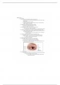
A. Bony orbital cavity surrounded by cushion of fat protects eye
1. Eyelids are like two movable shades that further protect eye from injury, strong
light, and dust
2. Upper eyelid larger and more mobile
3. Eyelashes are short hairs in double or triple rows that curve outward from lid
margins, filtering out dust and dirt
B. Palpebral fissure: elliptical open space between eyelids
1. When closed, lid margins approximate completely
2. When open, upper lid covers part of iris
C. Lower lid margin, at limbus, borders between cornea and sclera
1. Canthus: corner of eye, angle where lids meet
a) Inner canthus: caruncle is small fleshy mass containing sebaceous glands
D. Tarsal plates : Within upper lid, they are strips of connective tissue that give it shape
1. Contain meibomian glands, which are modified sebaceous glands that secrete an
oily lubricating material onto lids
a) This stops the tears from overflowing and helps to form an airtight seal
when lids are closed
E. Conjunctiva: transparent protective covering of exposed part of eye
1. Palpebral conjunctiva: lines lids, is clear, with many small blood vessels
2. Bulbar conjunctiva: overlies eyeball, with white sclera showing through
a) At limbus, conjunctivae merge with cornea
F. Cornea: covers and protects iris and pupil
G. Lacrimal gland, in upper outer corner over eye, secretes tears
1. Tears wash across eye and drawn up evenly as lid blinks
2. Drain into puncta, on upper and lower lids at inner canthus
3. Then drain into nasolacrimal sac, through ½-inch-long nasolacrimal duct, and
empty into inferior meatus inside nose
4.
H. Extraocular Muscles
1. Six muscles attach eyeball to its orbit and direct eye to points of person’s interest
a) Give eye both straight and rotary movement
(1) Four straight, or rectus, muscles are superior, inferior, lateral,
and medial rectus muscles
I. External Anatomy
A. Bony orbital cavity surrounded by cushion of fat protects eye
1. Eyelids are like two movable shades that further protect eye from injury, strong
light, and dust
2. Upper eyelid larger and more mobile
3. Eyelashes are short hairs in double or triple rows that curve outward from lid
margins, filtering out dust and dirt
B. Palpebral fissure: elliptical open space between eyelids
1. When closed, lid margins approximate completely
2. When open, upper lid covers part of iris
C. Lower lid margin, at limbus, borders between cornea and sclera
1. Canthus: corner of eye, angle where lids meet
a) Inner canthus: caruncle is small fleshy mass containing sebaceous glands
D. Tarsal plates : Within upper lid, they are strips of connective tissue that give it shape
1. Contain meibomian glands, which are modified sebaceous glands that secrete an
oily lubricating material onto lids
a) This stops the tears from overflowing and helps to form an airtight seal
when lids are closed
E. Conjunctiva: transparent protective covering of exposed part of eye
1. Palpebral conjunctiva: lines lids, is clear, with many small blood vessels
2. Bulbar conjunctiva: overlies eyeball, with white sclera showing through
a) At limbus, conjunctivae merge with cornea
F. Cornea: covers and protects iris and pupil
G. Lacrimal gland, in upper outer corner over eye, secretes tears
1. Tears wash across eye and drawn up evenly as lid blinks
2. Drain into puncta, on upper and lower lids at inner canthus
3. Then drain into nasolacrimal sac, through ½-inch-long nasolacrimal duct, and
empty into inferior meatus inside nose
4.
H. Extraocular Muscles
1. Six muscles attach eyeball to its orbit and direct eye to points of person’s interest
a) Give eye both straight and rotary movement
(1) Four straight, or rectus, muscles are superior, inferior, lateral,
and medial rectus muscles