Lecture notes Anatomy and Physiology
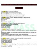
This document, titled "Wall of the GIT", offers a detailed explanation of the structure and functional anatomy of the gastrointestinal tract (GIT), specifically focusing on the various layers of the GIT and the salivary glands. Here's an in-depth explanation: Wall of the GIT: The GIT wall consists of four layers, and each one plays a crucial role in digestion and absorption. These layers are detailed as follows: 1. Mucosa: Location: Innermost layer of the GIT. Function: Secretes mucus, hormones, and enzymes; involved in absorption and protection. Components: Epithelium: In direct contact with food, responsible for secretion and absorption (e.g., simple columnar epithelium and stratified squamous epithelium in specific regions). Lamina propria: Loose connective tissue containing blood vessels and lymphatic tissue for nutrient absorption and immune defense. Muscularis mucosa: A thin layer of smooth muscle that enhances the mucosa's ability to absorb nutrients by causing local movements. 2. Submucosa: Description: The second layer of the GIT wall. Function: Contains blood vessels, lymphatics, and nerves. Components: Submucosal (Meissner's) plexus: Plays a role in controlling secretions and blood flow to the digestive organs. This layer works independently of the central nervous system (CNS) for some digestive functions. 3. Muscularis Externa: Description: The third layer, consisting of two smooth muscle layers (inner circular layer and outer longitudinal layer). Function: Responsible for propulsion and segmentation (breaking down food). Components: Peristalsis: Involuntary contractions that propel food along the digestive tract. Segmentation: Localized contractions that mix food with digestive enzymes. Nerve plexus: Myenteric (Auerbach's) plexus coordinates the muscle activity. 4. Serosa: Description: The outermost layer. Function: Protective layer that reduces friction as the digestive organs move and contracts. Components: It is a thin, slippery membrane consisting of simple squamous epithelium and a thin layer of areolar connective tissue. In areas not covered by the serosa, the adventitia (dense connective tissue) provides support. Functional Anatomy of the GI Tract: This section highlights the role and function of the GI tract, particularly focusing on digestion, secretion, absorption, and the structure of its layers, as described above. Salivary Glands: This part of the document focuses on the salivary glands, their functions, and the components of saliva. Definition: Glands associated with the oral cavity that secrete saliva. Functions of Saliva: Cleanses the mouth: Helps remove food particles and bacteria. Dissolves food chemicals: So they can be tasted. Moistens food: Helps in forming a bolus (soft mass of chewed food) for easier swallowing. Contains the enzyme amylase: Begins the digestion of starch (complex carbohydrates). Components of Saliva: 97-99.5% water: Keeps the oral cavity moist and lubricates food. Electrolytes: Includes ions such as Na+, K+, PO4^3-, and HCO3-. Digestive enzymes: Salivary amylase: Begins carbohydrate digestion in the mouth. Lingual lipase: Begins the digestion of fats but becomes more active in the stomach. Mucins: Glycoproteins that lubricate and protect the oral cavity and the food. This document provides a detailed breakdown of the GIT's structure and the role of the salivary glands, organized in a concise and clear way. Important functions and processes are written with emphasis using color-coded text (black, blue, and red), making the content easy to navigate and understand. This can serve as a useful study guide for students learning about the digestive system, focusing on both the structural and functional aspects of the gastrointestinal tract.
Written for
- Institution
- Tshwane University of Technology (TUT)
- Course
- Anatomy and Physiology
Document information
- Uploaded on
- September 15, 2024
- Number of pages
- Unknown
- Written in
- 2024/2025
- Type
- Class notes
- Professor(s)
- Dr maimele
- Contains
- All classes
Subjects
-
saliva