MUST KNOW THIS notes and images for exam 05-12-19 200300074
KEY ITEMS of Cognitive
Neuroscience
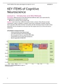
Lecture 1 – Introduction and EEG Methods
Phrenology: different areas on the skull represent different skills, each represented by
enlargements or indentations of the skull
Start for anatomy (Franz Joseph Gall)
Neurons: each neuron has a different function (motor, sensory), they all have an input
(=dendrites), output (=synapse), a transfer (=axon) and a modulator (=myelin). Neurons
don’t reproduce after birth, but their connections do alter (learning). Neuron transfer action
potentials from the dendrites to the axons into the synapse and so on to the next neuron
(dendrites).
All methods to measure the brain
- Action potentials (electrophysiology)
- Local field potentials (electrophysiology)
- Electromagnetic fields at scalp (EEG/ERP/MEG)
- Manipulating neural activity (perturbations)
- Blood oxygenation (fMRI/PET)
1
, MUST KNOW THIS notes and images for exam 05-12-19 200300074
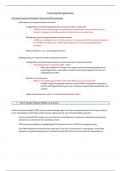
ElectroEncephaloGraphy – EEG
Measures differences in voltage, based on
local field potential, across the scalp
Reflects the post-synaptic potentials of
both inhibitory and excitatory potentials =
input
Measures mainly activity in gyri
Great temporal resolution, okay spatial
resolution
MagnetoEncephaloGraphy – MEG
Measures the magnetic field with a better
localization than EEG
Measures only activity from sulci
Expensive
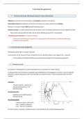
Brain wave states
Slow waves (e.g. theta) = low arousal
Fast waves (e.g. beta) = high arousal
Each frequency reflects a different arousal state
o Gamma (32Hz>) = superlearning
o Beta (16-31Hz) = processing information, analytical thinking
o Alpha (8-15Hz) = Eyes closed or very relaxed
o Theta (4-7Hz) = Sleep, REM, dreaming, deep meditation
o Delta (<4Hz) = Deep dreamless sleep
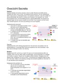
Event-related potentials (ERP)
On the background of EEG waves
Noise-free average
Fine changes in response to stimuli
Measures peaks and troughs (amount of activity and timing of activity) Distribution
of peak (or
trough) across
the scalp by
multiple
electrodes and
looking at
differences
between those
across scalp
2
KEY ITEMS of Cognitive
Neuroscience
Lecture 1 – Introduction and EEG Methods
Phrenology: different areas on the skull represent different skills, each represented by
enlargements or indentations of the skull
Start for anatomy (Franz Joseph Gall)
Neurons: each neuron has a different function (motor, sensory), they all have an input
(=dendrites), output (=synapse), a transfer (=axon) and a modulator (=myelin). Neurons
don’t reproduce after birth, but their connections do alter (learning). Neuron transfer action
potentials from the dendrites to the axons into the synapse and so on to the next neuron
(dendrites).
All methods to measure the brain
- Action potentials (electrophysiology)
- Local field potentials (electrophysiology)
- Electromagnetic fields at scalp (EEG/ERP/MEG)
- Manipulating neural activity (perturbations)
- Blood oxygenation (fMRI/PET)
1
, MUST KNOW THIS notes and images for exam 05-12-19 200300074
ElectroEncephaloGraphy – EEG
Measures differences in voltage, based on
local field potential, across the scalp
Reflects the post-synaptic potentials of
both inhibitory and excitatory potentials =
input
Measures mainly activity in gyri
Great temporal resolution, okay spatial
resolution
MagnetoEncephaloGraphy – MEG
Measures the magnetic field with a better
localization than EEG
Measures only activity from sulci
Expensive
Brain wave states
Slow waves (e.g. theta) = low arousal
Fast waves (e.g. beta) = high arousal
Each frequency reflects a different arousal state
o Gamma (32Hz>) = superlearning
o Beta (16-31Hz) = processing information, analytical thinking
o Alpha (8-15Hz) = Eyes closed or very relaxed
o Theta (4-7Hz) = Sleep, REM, dreaming, deep meditation
o Delta (<4Hz) = Deep dreamless sleep
Event-related potentials (ERP)
On the background of EEG waves
Noise-free average
Fine changes in response to stimuli
Measures peaks and troughs (amount of activity and timing of activity) Distribution
of peak (or
trough) across
the scalp by
multiple
electrodes and
looking at
differences
between those
across scalp
2