Lectures Systems
Neuroanatomy
Second motorneuron
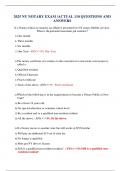
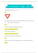
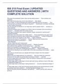
First motor neuron is the upper motor neuron and is in the motor cortex. Pyramidal
projection. Connects to the second motor neuron. Second motor neuron is with
alpha motor neurons. Is the lower motor neuron. Is in the brainstem or the ventral
horn of the spinal cord. Connects to the striated muscles.
The cortex controls the first motor neurons. The second motor neurons control
muscle.
Each skeletal muscle consists of striated muscle fibers. Reach from tendon to
tendon. Muscles works over a joint. There are agonistic muscles that work
together (like two flexors) and antagonist muscles that work against each other (a
flexor and an extensor). All muscles in an extremity have to work together.
For smooth movement there must be a very close cooperation between muscles
that act on a specific joint. Sherrington’s law of reciprocal innervation; contraction of an
agonist requires relaxation of antagonist. The cooperation of muscles is difficult, so the basal
ganglia and the cerebellar subsystems work together.
Each muscle fiber is innervated by a single axon. This axon originates from an alpha motor
neuron which is also called the second motor neuron or the lower motor neuron. They are
located in the ventral horn of the spinal cord.
Between the alpha motor neuron and the muscle fiber is a large specialized synapse; the
motor end plate/neuromuscular junction. Neurotransmitter is acetylcholine. Motor endplate
is a very effective synapse. Every neuronal action potential will cause an action potential in
the muscle fiber. This is because of the large surface (invaginations) and the high density of
postsynaptic receptors. A muscle fiber AP results in a twitch. Release of calcium will lead to
shoving of the fibers over each other and thus contraction. Calcium is continuously removed.
Calcium is only shortly available. Next muscle fiber AP can already be evoked before the
muscle fiber twitch is over. This because a twitch lasts 25-200ms and the AP only 5ms. The
more APs you send, the more contraction you get. Frequency of APs determines the
contraction. A train of APs will tetanize the muscle; fused, complete contraction.
Extensor muscles of the back are stronger because this is antigravity. Antigravity muscles
are always stronger.
,The frequency of the alpha motor neuron AP determines the amount of contraction. One
alpha motor neuron may innervate many muscle fibers at the same time. A motor unit is the
alpha motor neuron together with all the muscle fibers that it innervates. Small units give a
more precise control of movement. Can control fibers more individually then.
The motor neuron pool is the group of motor neurons that together innervate all the muscle
fibers of a single complete muscle. They form a group together in the ventral horn.
Neighborhood relations in the muscles is reflected in the spinal cord.
In the spinal column there are two separate areas for the motor neurons. The medial
somatomotor column is all over the entire length of the spinal cord and is for postural
musculature. The lateral somatomotor column is only in the intumescences and is for the
limb musculature.
Size principle of motor neuron recruitment
Size principle of Henneman is the size principle of motor neuron recruitment.
There is a direct relation between the size of the neuron (soma) and the surface of cell
membrane it has to maintain. Long axons, large dendritic tree, many branches and a thick
axons requires a large soma. More to take care of. Large neuron is more difficult to excite
than a small neuron.
Smaller motor units allow for more precise control of muscle contraction. The size of a motor
neuron is proportional to number of muscle fibers it innervates. Large motor unit, large motor
neuron. The smaller a motor neuron is, the easier it is excited. Less energetic firing will lead
to first action of the small motor neurons and slow contractions. More energy (higher
frequency of action potentials), larger motor units that react. So; the increase of the
frequency of the input into the motor neuron pool results in increasing force exerted by the
muscle. If there is an increase in the drive, automatically motor neurons from small to large
will be recruited. As a consequence also the motor units will be recruited from small to
large. The size principle is very efficient, but there is only forward control and no feedback.
There are also different types of muscle fibers. Dark fibers are slow, light fibers are fast. All
fibers in a motor unit are of the same type. The motor neuron dictates the muscle fiber type.
Small motor units with small motor neurons innervate slow but fatigue resistant muscle
fibers. The large motor units are large and fast but fatigue easily. So; there is also a relation
between the motor neuron size and the muscle fiber type.
Progressive increase of drive automatically recruits the motor neurons from small to
large
Consequently progressive increase of drive automatically recruits the motor units
from small to large
Increasing the drive results in progressive increase of the force exerted by the muscle
Feedback circuits
Stretch sensor parallel to the muscle fibers. Is called the muscle spindle. Biosensor only
works when it virtually has the same length all the time. Is then very sensitive. Cannot stand
large deformations. To make this possible, the stretch sensor is attached to an intrafusal
muscle fiber. This muscle fiber will adapt the length of the stretch sensor to changes in the
length of the entire muscle. At a given length of the muscle the sensitivity of the stretch
sensor can be controlled through this muscle fiber. Is only if you actively yourself use the
muscle. This to only sense when there is external influence of the muscle stretch.
,Both tonic and phasic sensors. Tonic; a signal as long as there is a change. As long as the
stretch lasts. Phasic is only when there is change. Signal stops when there is no change,
adapts to the new situation.
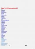
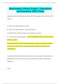
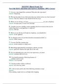
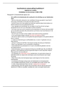
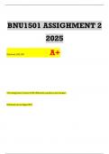
The intrafusal muscle fiber is innervated by a
gamma motor neuron that is also part of the
motor neuron pool. It controls the length and
thus the sensitivity of the stretch sensor.
Separate gamma motor neurons for tonic and
phasic stretch sensors. Gamma motor neurons
are really small. Alpha motor neurons are way
bigger. In a voluntary movement, the alpha
and gamma motor neurons are activated at the
same time so there will be no change of
signal during a voluntary movement.
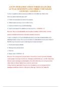
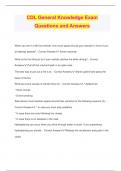
Stretch of the stretch sensor by an external
force will lead to an increase in the drive of all
motor neurons in the motor neuron in the pool.
This muscle will shorten, the sensor
unstretches and the drive will eventually diminishes. The stretch sensor
activates the alpha motor neurons. Not the gamma motor neuron too prevent
tetanization of the muscle; the signal by the spindle would only increase. A
reflex circuit (myotatic reflex) to remain the muscle length in the case of
external forces. Is monosynaptic;
very fast. If there is pre-stretch, the
spindle is more sensitive. The
myotatic reflex is also called the
gamma loop. It maintains the
length of muscles upon disturbance
by an external factor like sudden
postural changes and change in
muscle load.
However, there must be
compensation by relaxation of the
antagonist. The antagonist muscle
is inhibited through an inhibitory
interneuron.
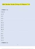
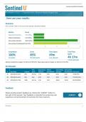
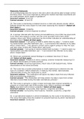
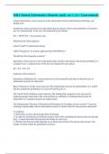
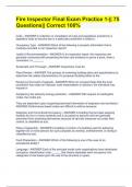
A second feedback system that is in series with the muscle fibers. There is also a stretch
sensor in tendon; Golgi tendon organ. Is a protective reflex; the inverse myotatic
reflex. Prevents the muscle from too much force that it will rip from the bone. Is not
monosynaptic. If the sensor
stretches, there is decrease of the
drive of all motor neuron in the
pool through an inhibitory
interneuron. The muscle will
lengthen, the sensor unstretches
and the inhibition will decrease
again. However, again because of
Sherington’s law the antagonist
will now be excited through an
, excitatory interneuron. Inverse myotatic reflex to protect the tendon and muscles and to
maintain the force and tone of the muscle. It is less sensitive than the myotatic reflex.
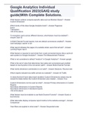
The myotatic reflex is monosynaptic and will make
the agonist contract. The stretch sensor is in parallel
and the circuit is there to maintain length of a muscle.
The inverse myotatic reflex is bisynaptic and makes
the agonist relax. The stretch sensor is in series and
the circuit is to maintain the force and tone of a
muscle. Both influence the antagonist as well
(reciprocal innervation)
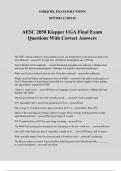
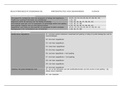
Another is pain sensor that gives feedback. Pain
sensor is in the epidermis and dermis in the skin.
Free nerve endings equipped with nociceptors. Are
chemical, thermal, chemical and polymodal.
However, in a pain reflex there must also be
compensation for the withdrawal of an extremity. Is
the nociceptive reflex. Is multisynaptic and
multisegmental. To really make sure that there is
withdrawal of the limb, there is also a feedback loop
that inhibits all the other reflexes. Happens
something to the other limb as well to support more.
There is ipsilateral flexion and contralateral
extension.
The whole circuit makes it possible for one single local neuron to control flexion in one leg
and simultaneously extension of the other leg. This is just a consequence of the assembly of
reflex circuits. More simple control of movement. Is more efficient (not so much large axons
from the cortex needed) and more easily coordinated, there is more cooperation.
An oscillatory circuit makes the control even more efficient. A tonic input will lead to a
oscillatory output. This allows bilateral coordination of the legs by one cortical neuron; in one
leg it will lead to flexion and in the other leg to extension. All rhythmic movements are
generated in the spinal cord; the central pattern generator.
Neuroanatomy
Second motorneuron
First motor neuron is the upper motor neuron and is in the motor cortex. Pyramidal
projection. Connects to the second motor neuron. Second motor neuron is with
alpha motor neurons. Is the lower motor neuron. Is in the brainstem or the ventral
horn of the spinal cord. Connects to the striated muscles.
The cortex controls the first motor neurons. The second motor neurons control
muscle.
Each skeletal muscle consists of striated muscle fibers. Reach from tendon to
tendon. Muscles works over a joint. There are agonistic muscles that work
together (like two flexors) and antagonist muscles that work against each other (a
flexor and an extensor). All muscles in an extremity have to work together.
For smooth movement there must be a very close cooperation between muscles
that act on a specific joint. Sherrington’s law of reciprocal innervation; contraction of an
agonist requires relaxation of antagonist. The cooperation of muscles is difficult, so the basal
ganglia and the cerebellar subsystems work together.
Each muscle fiber is innervated by a single axon. This axon originates from an alpha motor
neuron which is also called the second motor neuron or the lower motor neuron. They are
located in the ventral horn of the spinal cord.
Between the alpha motor neuron and the muscle fiber is a large specialized synapse; the
motor end plate/neuromuscular junction. Neurotransmitter is acetylcholine. Motor endplate
is a very effective synapse. Every neuronal action potential will cause an action potential in
the muscle fiber. This is because of the large surface (invaginations) and the high density of
postsynaptic receptors. A muscle fiber AP results in a twitch. Release of calcium will lead to
shoving of the fibers over each other and thus contraction. Calcium is continuously removed.
Calcium is only shortly available. Next muscle fiber AP can already be evoked before the
muscle fiber twitch is over. This because a twitch lasts 25-200ms and the AP only 5ms. The
more APs you send, the more contraction you get. Frequency of APs determines the
contraction. A train of APs will tetanize the muscle; fused, complete contraction.
Extensor muscles of the back are stronger because this is antigravity. Antigravity muscles
are always stronger.
,The frequency of the alpha motor neuron AP determines the amount of contraction. One
alpha motor neuron may innervate many muscle fibers at the same time. A motor unit is the
alpha motor neuron together with all the muscle fibers that it innervates. Small units give a
more precise control of movement. Can control fibers more individually then.
The motor neuron pool is the group of motor neurons that together innervate all the muscle
fibers of a single complete muscle. They form a group together in the ventral horn.
Neighborhood relations in the muscles is reflected in the spinal cord.
In the spinal column there are two separate areas for the motor neurons. The medial
somatomotor column is all over the entire length of the spinal cord and is for postural
musculature. The lateral somatomotor column is only in the intumescences and is for the
limb musculature.
Size principle of motor neuron recruitment
Size principle of Henneman is the size principle of motor neuron recruitment.
There is a direct relation between the size of the neuron (soma) and the surface of cell
membrane it has to maintain. Long axons, large dendritic tree, many branches and a thick
axons requires a large soma. More to take care of. Large neuron is more difficult to excite
than a small neuron.
Smaller motor units allow for more precise control of muscle contraction. The size of a motor
neuron is proportional to number of muscle fibers it innervates. Large motor unit, large motor
neuron. The smaller a motor neuron is, the easier it is excited. Less energetic firing will lead
to first action of the small motor neurons and slow contractions. More energy (higher
frequency of action potentials), larger motor units that react. So; the increase of the
frequency of the input into the motor neuron pool results in increasing force exerted by the
muscle. If there is an increase in the drive, automatically motor neurons from small to large
will be recruited. As a consequence also the motor units will be recruited from small to
large. The size principle is very efficient, but there is only forward control and no feedback.
There are also different types of muscle fibers. Dark fibers are slow, light fibers are fast. All
fibers in a motor unit are of the same type. The motor neuron dictates the muscle fiber type.
Small motor units with small motor neurons innervate slow but fatigue resistant muscle
fibers. The large motor units are large and fast but fatigue easily. So; there is also a relation
between the motor neuron size and the muscle fiber type.
Progressive increase of drive automatically recruits the motor neurons from small to
large
Consequently progressive increase of drive automatically recruits the motor units
from small to large
Increasing the drive results in progressive increase of the force exerted by the muscle
Feedback circuits
Stretch sensor parallel to the muscle fibers. Is called the muscle spindle. Biosensor only
works when it virtually has the same length all the time. Is then very sensitive. Cannot stand
large deformations. To make this possible, the stretch sensor is attached to an intrafusal
muscle fiber. This muscle fiber will adapt the length of the stretch sensor to changes in the
length of the entire muscle. At a given length of the muscle the sensitivity of the stretch
sensor can be controlled through this muscle fiber. Is only if you actively yourself use the
muscle. This to only sense when there is external influence of the muscle stretch.
,Both tonic and phasic sensors. Tonic; a signal as long as there is a change. As long as the
stretch lasts. Phasic is only when there is change. Signal stops when there is no change,
adapts to the new situation.
The intrafusal muscle fiber is innervated by a
gamma motor neuron that is also part of the
motor neuron pool. It controls the length and
thus the sensitivity of the stretch sensor.
Separate gamma motor neurons for tonic and
phasic stretch sensors. Gamma motor neurons
are really small. Alpha motor neurons are way
bigger. In a voluntary movement, the alpha
and gamma motor neurons are activated at the
same time so there will be no change of
signal during a voluntary movement.
Stretch of the stretch sensor by an external
force will lead to an increase in the drive of all
motor neurons in the motor neuron in the pool.
This muscle will shorten, the sensor
unstretches and the drive will eventually diminishes. The stretch sensor
activates the alpha motor neurons. Not the gamma motor neuron too prevent
tetanization of the muscle; the signal by the spindle would only increase. A
reflex circuit (myotatic reflex) to remain the muscle length in the case of
external forces. Is monosynaptic;
very fast. If there is pre-stretch, the
spindle is more sensitive. The
myotatic reflex is also called the
gamma loop. It maintains the
length of muscles upon disturbance
by an external factor like sudden
postural changes and change in
muscle load.
However, there must be
compensation by relaxation of the
antagonist. The antagonist muscle
is inhibited through an inhibitory
interneuron.
A second feedback system that is in series with the muscle fibers. There is also a stretch
sensor in tendon; Golgi tendon organ. Is a protective reflex; the inverse myotatic
reflex. Prevents the muscle from too much force that it will rip from the bone. Is not
monosynaptic. If the sensor
stretches, there is decrease of the
drive of all motor neuron in the
pool through an inhibitory
interneuron. The muscle will
lengthen, the sensor unstretches
and the inhibition will decrease
again. However, again because of
Sherington’s law the antagonist
will now be excited through an
, excitatory interneuron. Inverse myotatic reflex to protect the tendon and muscles and to
maintain the force and tone of the muscle. It is less sensitive than the myotatic reflex.
The myotatic reflex is monosynaptic and will make
the agonist contract. The stretch sensor is in parallel
and the circuit is there to maintain length of a muscle.
The inverse myotatic reflex is bisynaptic and makes
the agonist relax. The stretch sensor is in series and
the circuit is to maintain the force and tone of a
muscle. Both influence the antagonist as well
(reciprocal innervation)
Another is pain sensor that gives feedback. Pain
sensor is in the epidermis and dermis in the skin.
Free nerve endings equipped with nociceptors. Are
chemical, thermal, chemical and polymodal.
However, in a pain reflex there must also be
compensation for the withdrawal of an extremity. Is
the nociceptive reflex. Is multisynaptic and
multisegmental. To really make sure that there is
withdrawal of the limb, there is also a feedback loop
that inhibits all the other reflexes. Happens
something to the other limb as well to support more.
There is ipsilateral flexion and contralateral
extension.
The whole circuit makes it possible for one single local neuron to control flexion in one leg
and simultaneously extension of the other leg. This is just a consequence of the assembly of
reflex circuits. More simple control of movement. Is more efficient (not so much large axons
from the cortex needed) and more easily coordinated, there is more cooperation.
An oscillatory circuit makes the control even more efficient. A tonic input will lead to a
oscillatory output. This allows bilateral coordination of the legs by one cortical neuron; in one
leg it will lead to flexion and in the other leg to extension. All rhythmic movements are
generated in the spinal cord; the central pattern generator.