Dr. S. Chakraborty
Skeletal System
The human body contains 206 bones.
Functions: major functions.
Movement Maintain or change position of body parts by
interacting with skeletal muscles.
Protection Enclose and protect the brain, lungs, and other organs
Support Support and anchor muscles.
Production Site for RBC and other blood cells.
Electrolyte balance Calcium and Phosphate ions
Storage Store mineral ions. In addition, the bone also store
energy reserve as lipids (known as yellow marrow).
Classification:
According to their location: 1) Axial skeleton and 2)Appendicular skeleton
Axial skeleton: The bones that form the body’s central axis. This includes skull, vertebral column
and the rib cage.
Appendicular skeleton: this includes upper and lower appendages, as well as bones used to attach
them to the axial skeleton.
According to their shape: 6 broad categories:
Long Short flat Irregular Sesamoid Sutural
Arm and Carpal bones Roof of Vertebrae, Found in Located
forearm, (wrist) and skull, the the pelvis joints of between the
thigh and tarsal sternum, the and several hand, knees flat bones of
leg, palms, bones(ankles) ribs, and the skull bones and feet. skull.**
soles, finger scapula (ethmoid, Patellae are
and toes. sphenoid the common
Longest etc) one. *
bone- femur
* Their location and abundance varies and this account for the disparities in total number of bones
sometime.
** There are 4 types of major sutures: 1) Lambdoid suture(separates two parietal and occipital bones
in skull); 2) Coronal suture (frontal to the parietal bones of either side); 3) Sagittal suture (between
the two parietal bones); 4)Squamous suture (between the parietal and temporal bones).
Bone structure:
Flat bones:
Flat bones like those of the cranium or the scapula are sandwiches of spongy bone between two
layers of compact bone. They are usually curved, so we can refer to an inner and outer table with
diploe between them. These diploe, especially in the skull, may become pneumatised, i.e. filled with
air. A ring of facial sinuses around the nose may become infected, leading to sinusitis.
Long Bone Structure:
Compact/Dense Bone: forms the bulk of the diaphyses of long bones; covers the spongy bone (the
epiphyses); its dense structure gives strength and rigidity to bones
Spongy Bone: makes up most of the tissue of the epiphyses of long bones
1
, Dr. S. Chakraborty
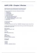
Diaphysis or Shaft: shaft/main portion of a long bone; contains space
called the medullary (marrow) cavity which contains fatty yellow
marrow
Endosteum: membrane that lines the medullary cavity; consists of a
single layer of osteoprogenitor cells
Epiphyses: the ends of long bones; contains red bone marrow, where
blood cell production (hemopoiesis) occurs
Epiphyseal Line: a bony structure that replaces the epiphyseal cartilage;
when it appears, the bone stops growing
Medullary Cavity (Marrow Cavity): a space within the diaphyses of long
bones; contains yellow marrow (stores fat)
Metaphysis: the region between the diaphyses and the epiphyses where
calcified matrix is replaced by bone; bone grows in length as a result of
this activity
Periosteum: a membrane that covers the outer surface of a bone; it has 2
layers: Fibrous Layer (outer layer that consists of fibroblasts and
collagen fibers)
Osteogenic Layer (contains osteoprogenitor cells)
Articular Cartilage: a thin layer of cartilage that covers the epiphyses of
a long bone; it reduces friction and absorbs shock at freely moveable
joints.
Microscopic structure of bones:
Compact Bones:
Structure: Haversian Systems (osteons) are the basic structural units of compact bone. Haversian
Sytems are located in the diaphyses of long bones; it covers spongy bone in the epiphyses (in short,
flat, or irregular bones it lies over the spongy bone). The functions of Haversian Systems are to
protect, support, and resist stress. Each system consists of the following:
1) Central Canal- a longitudinal canal; contains blood vessels and nerves
2) Volkmann’s canal or perforating canal or horizontal canal
3) Lamellae- concentric rings of hard, calcified matrix surrounding central canals
4) Lacunae- small spaces between the lamellae; each lacuna contains one osteocyte
5) Osteocytes- mature bone cells that maintain the bony matrix
6) Canaliculi- minute channels that house the filopodial process of osteocytes
Bone Cells (widely separated throughout the matrix)
1) Osteoprogenitor Cells:
function- divide by mitosis and develop into osteoblasts
location- inner layer of periosteum; endosteum; central and perforating canals
2) Osteoblasts (cells derived from)
function- form bone tissue by secreting matrix (collagen + other organic compounds)
location: surface of bones.
2
Skeletal System
The human body contains 206 bones.
Functions: major functions.
Movement Maintain or change position of body parts by
interacting with skeletal muscles.
Protection Enclose and protect the brain, lungs, and other organs
Support Support and anchor muscles.
Production Site for RBC and other blood cells.
Electrolyte balance Calcium and Phosphate ions
Storage Store mineral ions. In addition, the bone also store
energy reserve as lipids (known as yellow marrow).
Classification:
According to their location: 1) Axial skeleton and 2)Appendicular skeleton
Axial skeleton: The bones that form the body’s central axis. This includes skull, vertebral column
and the rib cage.
Appendicular skeleton: this includes upper and lower appendages, as well as bones used to attach
them to the axial skeleton.
According to their shape: 6 broad categories:
Long Short flat Irregular Sesamoid Sutural
Arm and Carpal bones Roof of Vertebrae, Found in Located
forearm, (wrist) and skull, the the pelvis joints of between the
thigh and tarsal sternum, the and several hand, knees flat bones of
leg, palms, bones(ankles) ribs, and the skull bones and feet. skull.**
soles, finger scapula (ethmoid, Patellae are
and toes. sphenoid the common
Longest etc) one. *
bone- femur
* Their location and abundance varies and this account for the disparities in total number of bones
sometime.
** There are 4 types of major sutures: 1) Lambdoid suture(separates two parietal and occipital bones
in skull); 2) Coronal suture (frontal to the parietal bones of either side); 3) Sagittal suture (between
the two parietal bones); 4)Squamous suture (between the parietal and temporal bones).
Bone structure:
Flat bones:
Flat bones like those of the cranium or the scapula are sandwiches of spongy bone between two
layers of compact bone. They are usually curved, so we can refer to an inner and outer table with
diploe between them. These diploe, especially in the skull, may become pneumatised, i.e. filled with
air. A ring of facial sinuses around the nose may become infected, leading to sinusitis.
Long Bone Structure:
Compact/Dense Bone: forms the bulk of the diaphyses of long bones; covers the spongy bone (the
epiphyses); its dense structure gives strength and rigidity to bones
Spongy Bone: makes up most of the tissue of the epiphyses of long bones
1
, Dr. S. Chakraborty
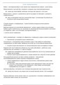
Diaphysis or Shaft: shaft/main portion of a long bone; contains space
called the medullary (marrow) cavity which contains fatty yellow
marrow
Endosteum: membrane that lines the medullary cavity; consists of a
single layer of osteoprogenitor cells
Epiphyses: the ends of long bones; contains red bone marrow, where
blood cell production (hemopoiesis) occurs
Epiphyseal Line: a bony structure that replaces the epiphyseal cartilage;
when it appears, the bone stops growing
Medullary Cavity (Marrow Cavity): a space within the diaphyses of long
bones; contains yellow marrow (stores fat)
Metaphysis: the region between the diaphyses and the epiphyses where
calcified matrix is replaced by bone; bone grows in length as a result of
this activity
Periosteum: a membrane that covers the outer surface of a bone; it has 2
layers: Fibrous Layer (outer layer that consists of fibroblasts and
collagen fibers)
Osteogenic Layer (contains osteoprogenitor cells)
Articular Cartilage: a thin layer of cartilage that covers the epiphyses of
a long bone; it reduces friction and absorbs shock at freely moveable
joints.
Microscopic structure of bones:
Compact Bones:
Structure: Haversian Systems (osteons) are the basic structural units of compact bone. Haversian
Sytems are located in the diaphyses of long bones; it covers spongy bone in the epiphyses (in short,
flat, or irregular bones it lies over the spongy bone). The functions of Haversian Systems are to
protect, support, and resist stress. Each system consists of the following:
1) Central Canal- a longitudinal canal; contains blood vessels and nerves
2) Volkmann’s canal or perforating canal or horizontal canal
3) Lamellae- concentric rings of hard, calcified matrix surrounding central canals
4) Lacunae- small spaces between the lamellae; each lacuna contains one osteocyte
5) Osteocytes- mature bone cells that maintain the bony matrix
6) Canaliculi- minute channels that house the filopodial process of osteocytes
Bone Cells (widely separated throughout the matrix)
1) Osteoprogenitor Cells:
function- divide by mitosis and develop into osteoblasts
location- inner layer of periosteum; endosteum; central and perforating canals
2) Osteoblasts (cells derived from)
function- form bone tissue by secreting matrix (collagen + other organic compounds)
location: surface of bones.
2