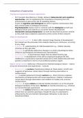
Class 7: genetics of Neurocutaneous
syndromes
Genetic mechanisms of heterozygous pathogenic
variants
- The genes implicated in Noonan syndrome and Noonan syndrome with
multiple lentigines are all part of the RAS-pathway and result mostly
from dominant activating variants causing increased signalling through
the RAS-MAPK pathway.
- A: homozyogous wild type allele in specific gene in all somatic
nucleated cells
- B: heterozygous pathogenic variant in specific gene in all somatic
nucleated cells
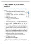
- Examples of cases of Noonan syndrome
o Most of the children have drooping eye lids
o Wide spead eyes
o Webbed neck
o …
- Many of the syndromes are caused by a heterozygous pathogenic variant in a
tumour suppressor gene with subsequent somatic inactivation of the normal
allele in aKected tissue
- PRKAR1A, NF1, SPRED1, NF2, SMARCB1, LZTR1, PTCH1, SUFU, PTEN, TSC1,
TSC2, VHL
- Autosomal dominant inheritance pattern but need for biallelic inactivation to
cause symptoms (recessive at the cellular level)
- A large intrafamilial variability in phenotypic expression is often seen because of
interindividual variability in the number, timing and location of second hits in wt
allele.
- The severity of the phenotype is often diKicult to predict from the genotype
- When you lose the other allele in the cell the cell starts to grow. Most of the
syndromes caused by a heterozygous pathogenic variant in the TSG are inherited
in an autosomal dominant way but if you look at the abnormal tissue you see a
second hit
o Whole list of conditions this occurs
- Each cell division have the risk of introducing a pathogenic variant
o in normal allele -> clump of cell that have both alleles inactivated
Café-au-lait spots
- Focal hyperpigmentation
- Always darker than surrounding skin
- Melanocytes with second hit in the NF1 gene
o Second hit is independent
- The size of the spot is correlated when the mutation occurs
- It shows that mutations are not rare, mutations in diving cells happen all the time
, TSC: clinical characteristics
- Little tumors at the level of the skin
- Other lesions at the level of the brain
o Second hit responsible for lesions
Nevoid basal cell carcinoma syndrome (NBCC)
- Basal cell carcinoma
o Usually don’t metastase
- Each of the tumors have a second hit
- A number of children develop a brain tumor
o Once they become older they disappear
Mosaicism
X-linked
- Epigenetic mosaicism due to inactivated x-chromosome
- The pathogenic variants in genes involved in the X-linked dominant
inherited syndromes are typically seen in females and are
embryonically lethal in males (IKBKG, NSDHL, HCCS, COX7B,
NDUFB11,OFD1)
- In a typical female with 2 X-chromosomes there is a patchwork with
cells where one X is inactivated
- There are females with 3 X chromosomes and then 2 are inactivated
- There are certain conditions know on the chromosome that are X-linked
dominant -> always expressed in females in a heterozygous situation
but at the same time are lethal in males
- Patchwork with normal X activated, and regions where the abnormal gene is
inactivated
- In a typical male there is only one X chromosome -> if the gene is present on that
X chromosome = lethal. There are a few exceptions
o Male embryos with Klinefelter syndrome (47,XXY)
o Embryos with a 46,XX male karyotype
o Embryos with post-zygotic mosaicism for the pathogenic variant
o A hypomorphic pathogenic variant associated with minimal activity of the
aKected protein suKicient for the cells to survive
- Example is Incontinentia Pigmenti
o Pathogenic variant in IKBKG (NF-KAPPA-B ESSENTIAL MODULATOR;
NEMO)
o Skin lesions in babies that disappear and leave only pigmentation marks
o Involvement of teeth, eyes and brain (epilepsy and intellectual disability)
o Hypomorphic allele in males cause immune deficiency syndrome
o In female half of the cell of the bone marrow contain a stemcell with the
functioning gene -> provide suKicient white blood cells -> avoid immune
deficiency syndrome
o Linear lesion -> region where the X-chromosome with the mutated gene is
active. They heal out -> after a couple of weeks you see scars
(hyperpigmented in the beginning but after a while with striped)
syndromes
Genetic mechanisms of heterozygous pathogenic
variants
- The genes implicated in Noonan syndrome and Noonan syndrome with
multiple lentigines are all part of the RAS-pathway and result mostly
from dominant activating variants causing increased signalling through
the RAS-MAPK pathway.
- A: homozyogous wild type allele in specific gene in all somatic
nucleated cells
- B: heterozygous pathogenic variant in specific gene in all somatic
nucleated cells
- Examples of cases of Noonan syndrome
o Most of the children have drooping eye lids
o Wide spead eyes
o Webbed neck
o …
- Many of the syndromes are caused by a heterozygous pathogenic variant in a
tumour suppressor gene with subsequent somatic inactivation of the normal
allele in aKected tissue
- PRKAR1A, NF1, SPRED1, NF2, SMARCB1, LZTR1, PTCH1, SUFU, PTEN, TSC1,
TSC2, VHL
- Autosomal dominant inheritance pattern but need for biallelic inactivation to
cause symptoms (recessive at the cellular level)
- A large intrafamilial variability in phenotypic expression is often seen because of
interindividual variability in the number, timing and location of second hits in wt
allele.
- The severity of the phenotype is often diKicult to predict from the genotype
- When you lose the other allele in the cell the cell starts to grow. Most of the
syndromes caused by a heterozygous pathogenic variant in the TSG are inherited
in an autosomal dominant way but if you look at the abnormal tissue you see a
second hit
o Whole list of conditions this occurs
- Each cell division have the risk of introducing a pathogenic variant
o in normal allele -> clump of cell that have both alleles inactivated
Café-au-lait spots
- Focal hyperpigmentation
- Always darker than surrounding skin
- Melanocytes with second hit in the NF1 gene
o Second hit is independent
- The size of the spot is correlated when the mutation occurs
- It shows that mutations are not rare, mutations in diving cells happen all the time
, TSC: clinical characteristics
- Little tumors at the level of the skin
- Other lesions at the level of the brain
o Second hit responsible for lesions
Nevoid basal cell carcinoma syndrome (NBCC)
- Basal cell carcinoma
o Usually don’t metastase
- Each of the tumors have a second hit
- A number of children develop a brain tumor
o Once they become older they disappear
Mosaicism
X-linked
- Epigenetic mosaicism due to inactivated x-chromosome
- The pathogenic variants in genes involved in the X-linked dominant
inherited syndromes are typically seen in females and are
embryonically lethal in males (IKBKG, NSDHL, HCCS, COX7B,
NDUFB11,OFD1)
- In a typical female with 2 X-chromosomes there is a patchwork with
cells where one X is inactivated
- There are females with 3 X chromosomes and then 2 are inactivated
- There are certain conditions know on the chromosome that are X-linked
dominant -> always expressed in females in a heterozygous situation
but at the same time are lethal in males
- Patchwork with normal X activated, and regions where the abnormal gene is
inactivated
- In a typical male there is only one X chromosome -> if the gene is present on that
X chromosome = lethal. There are a few exceptions
o Male embryos with Klinefelter syndrome (47,XXY)
o Embryos with a 46,XX male karyotype
o Embryos with post-zygotic mosaicism for the pathogenic variant
o A hypomorphic pathogenic variant associated with minimal activity of the
aKected protein suKicient for the cells to survive
- Example is Incontinentia Pigmenti
o Pathogenic variant in IKBKG (NF-KAPPA-B ESSENTIAL MODULATOR;
NEMO)
o Skin lesions in babies that disappear and leave only pigmentation marks
o Involvement of teeth, eyes and brain (epilepsy and intellectual disability)
o Hypomorphic allele in males cause immune deficiency syndrome
o In female half of the cell of the bone marrow contain a stemcell with the
functioning gene -> provide suKicient white blood cells -> avoid immune
deficiency syndrome
o Linear lesion -> region where the X-chromosome with the mutated gene is
active. They heal out -> after a couple of weeks you see scars
(hyperpigmented in the beginning but after a while with striped)