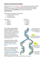
Chapter 2 Definitions:
instrument, uses visible light and lenses to magnify Light
objects microscope
LM that uses 2 lenses (objective & eyepiece) to magnify an Compound light
object microscope
using different stains to distinguish types of cells Differential
staining
application of 2nd stain to create contrast in colour Counterstain
how many times bigger that real life the object being Magnification
viewed is
shortest distance objects can be seen as separate Resolution
using a beam of electrons to illuminate a specimen Electron
microscopy
features of cells that can only be seen with an EM Ultrastructure
objects/structures on a microscope image that have been Artefacts
created by processing the specimen
beam of electrons transmitted through specimen to TEM
produce image
beam of electrons transmitted over surface of specimen to SEM
make 3D surface image
beam of fluorescence through pinhole to make image Laser scanning
confocal
microscope
cells with no membrane-bound nucleus/organelles Prokaryotic
single celled prokaryotic organisms Prokaryotes
cells with a nucleus and membrane-bound organelles Eukaryotic
multi/unicellular organisms with eukaryotic cells Eukaryotes
internal fluid of cell made of cytosol, organelles and Cytoplasm
cytoskeleton
membrane-bound compartments with varying functions in Organelles
eukaryotic cells
contains genetic material of the cell, controls Nucleus
cell functions
area within nucleus that produces ribosomes Nucleolus
proteins that form complex with DNA Histones
uncondensed DNA complex w/histones Chromatin
condensed DNA coils (chromatin), visible when cells divide Chromosomes