Part 2 - Video Lecture Notes
Thursday, February 7, 2019 11:08 AM
Separation Techniques Review
• SDS PAGE: electrophoresis gels of SDS-bound proteins
• 1 band is found on a PAGE:
○ Most likely 1 protein present, however it could also be more than one protein with the same MW.
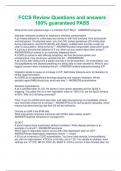
• Isoelectric focusing:
○ All proteins first inserted into the side with high pH (basic).
○ Proteins that are at pHs higher than their pI-> negatively charged.
○ Proteins will move down the strip in order to reach their pI, @ a neutral zero charge.
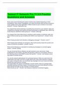
• Proteome: visual of all the proteins in a cell. Similar to genomes.
• Comparing proteomes thru 2D gels can help identify differences in healthy vs. cancerous tissues. Ex. One spot is darker in gel->
protein is being up-regulated in cancerous colon tissue vs healthy colon tissue.
Thursday, February 7, 2019 11:08 AM
Separation Techniques Review
• SDS PAGE: electrophoresis gels of SDS-bound proteins
• 1 band is found on a PAGE:
○ Most likely 1 protein present, however it could also be more than one protein with the same MW.
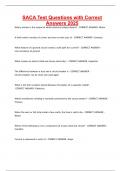
• Isoelectric focusing:
○ All proteins first inserted into the side with high pH (basic).
○ Proteins that are at pHs higher than their pI-> negatively charged.
○ Proteins will move down the strip in order to reach their pI, @ a neutral zero charge.
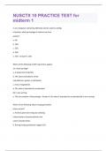
• Proteome: visual of all the proteins in a cell. Similar to genomes.
• Comparing proteomes thru 2D gels can help identify differences in healthy vs. cancerous tissues. Ex. One spot is darker in gel->
protein is being up-regulated in cancerous colon tissue vs healthy colon tissue.