Principles and Practice II
Chapter 31:
COLORECTAL:
Colorectal cancer is the second leading cause of cancer deaths in the U.S
Caused by:
- Diet high in animal fat and low in fiber
- Chronic ulcerative colitis (carcinomas in adenomatous polyps, usually occurs in the
rectum and sigmoid colon, intensive inflammation of the bowel wall and ulceration,
bloody diarrhea attacks up to 20 times a day)
o The risk of developing colorectal cancer from chronic ulcerative colitis depends
on the extent of bowel involvement, age of onset, and severity and duration of
disease)
o Adenomatous polyps (precursor to malignancy) are growths that arise from the
mucosal lining and protrude into the lumen of the bowel
- Hereditary cancer syndrome (FAP and HNPCC, aka Lynch syndrome)
o Almost all patients with FAP (familial adenomatous polyposis) develop colon
cancer if left untreated (FAP is treated with the complete removal of the colon and
rectum)
o Lynch syndrome is the most common form of hereditary colorectal cancer
syndrome (without polyposis)
o Gardner syndrome is similar to FAP, polyposis growths in the large bowel
- First degree relative with colorectal cancer
Major factor in determining treatment is area of malignancy (retroperitoneally or
intraperitoneally)
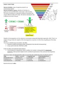
8 regions of the colon:
1) Cecum (I)
2) Ascending colon
3) Descending colon
4) Splenic flexure
5) Hepatic flexure
6) Transverse colon (I)
7) Sigmoid colon (I)
8) Rectum
(I=intraperitoneally) – complete mesentery and serosa, freely mobile (usually surgically
removable, if failure, peritoneal seeding indicated)
(others are located retroperitoneally)
Retroperitoneally located anatomy commonly spreads outside the bowel wall and invades
surrounding structures
, The rectum is continuous with the sigmoid and begins at S3
Upper rectum is covered by the peritoneum on its lateral and anterior surfaces
Transverse folds divide rectum into the upper valve, middle valve, and lower value (ampulla)
Mucosa – innermost layer, forms the lumen
Submucosa – rich in blood vessels and lymphatics
The lymphatic drainage of the colon follows the mesenteric vessels
Patients with rectal cancer usually have a change in bowel habits, diarrhea, change in stool
caliber, dark/black covered stools, tenesmus (rectal spasms), and/or rectal bleeding
(hematochezia)
Left colon: obstructive and abdominal pain
Right colon: abdominal pain and mass
Screening guidelines – 45 years of age
A colonoscopy examines the entire colon and visualizes polyps
- Should be done every 10 years
Patients with colorectal cancer should get a digital rectal examination
- Attention to the lesion size, location from the anal verge and rectal wall, and mobility
A proctosigmoidoscopy allows more accuracy than colonoscopy and determines whether the
mass is exophytic or ulcerative
Supraclavicular lymph node involvement indicates extensive incurable disease, result of
paraaortic nodes travelling via the thoracic duct
CBC (complete blood count) and blood chemistry profile is used for diagnosis
Carcinoembryonic antigen (CEA) is a protein molecule associated with colon and ovarian cancer
Adenocarcinoma is the most common malignancy of the large bowel (90%-95%)
AJCC TNM
Most important prognostic indicators of survival:
1) Lymph nodes
2) Depth of penetration through the bowel wall
Dukes staging system:
A – not penetrated bowel wall
B – penetrated bowel wall
C – penetrated bowel wall and positive nodes
Lymphatic spread occurs if the tumor has invaded the submucosal layer of the bowel
Blood-borne spread to the liver is the most common type of distant mets
- Involves the venous drainage of the GI system
Second most common site of distant mets spread is the lung (tumor in the IVC)
SURGERY IS TOC
Chapter 31:
COLORECTAL:
Colorectal cancer is the second leading cause of cancer deaths in the U.S
Caused by:
- Diet high in animal fat and low in fiber
- Chronic ulcerative colitis (carcinomas in adenomatous polyps, usually occurs in the
rectum and sigmoid colon, intensive inflammation of the bowel wall and ulceration,
bloody diarrhea attacks up to 20 times a day)
o The risk of developing colorectal cancer from chronic ulcerative colitis depends
on the extent of bowel involvement, age of onset, and severity and duration of
disease)
o Adenomatous polyps (precursor to malignancy) are growths that arise from the
mucosal lining and protrude into the lumen of the bowel
- Hereditary cancer syndrome (FAP and HNPCC, aka Lynch syndrome)
o Almost all patients with FAP (familial adenomatous polyposis) develop colon
cancer if left untreated (FAP is treated with the complete removal of the colon and
rectum)
o Lynch syndrome is the most common form of hereditary colorectal cancer
syndrome (without polyposis)
o Gardner syndrome is similar to FAP, polyposis growths in the large bowel
- First degree relative with colorectal cancer
Major factor in determining treatment is area of malignancy (retroperitoneally or
intraperitoneally)
8 regions of the colon:
1) Cecum (I)
2) Ascending colon
3) Descending colon
4) Splenic flexure
5) Hepatic flexure
6) Transverse colon (I)
7) Sigmoid colon (I)
8) Rectum
(I=intraperitoneally) – complete mesentery and serosa, freely mobile (usually surgically
removable, if failure, peritoneal seeding indicated)
(others are located retroperitoneally)
Retroperitoneally located anatomy commonly spreads outside the bowel wall and invades
surrounding structures
, The rectum is continuous with the sigmoid and begins at S3
Upper rectum is covered by the peritoneum on its lateral and anterior surfaces
Transverse folds divide rectum into the upper valve, middle valve, and lower value (ampulla)
Mucosa – innermost layer, forms the lumen
Submucosa – rich in blood vessels and lymphatics
The lymphatic drainage of the colon follows the mesenteric vessels
Patients with rectal cancer usually have a change in bowel habits, diarrhea, change in stool
caliber, dark/black covered stools, tenesmus (rectal spasms), and/or rectal bleeding
(hematochezia)
Left colon: obstructive and abdominal pain
Right colon: abdominal pain and mass
Screening guidelines – 45 years of age
A colonoscopy examines the entire colon and visualizes polyps
- Should be done every 10 years
Patients with colorectal cancer should get a digital rectal examination
- Attention to the lesion size, location from the anal verge and rectal wall, and mobility
A proctosigmoidoscopy allows more accuracy than colonoscopy and determines whether the
mass is exophytic or ulcerative
Supraclavicular lymph node involvement indicates extensive incurable disease, result of
paraaortic nodes travelling via the thoracic duct
CBC (complete blood count) and blood chemistry profile is used for diagnosis
Carcinoembryonic antigen (CEA) is a protein molecule associated with colon and ovarian cancer
Adenocarcinoma is the most common malignancy of the large bowel (90%-95%)
AJCC TNM
Most important prognostic indicators of survival:
1) Lymph nodes
2) Depth of penetration through the bowel wall
Dukes staging system:
A – not penetrated bowel wall
B – penetrated bowel wall
C – penetrated bowel wall and positive nodes
Lymphatic spread occurs if the tumor has invaded the submucosal layer of the bowel
Blood-borne spread to the liver is the most common type of distant mets
- Involves the venous drainage of the GI system
Second most common site of distant mets spread is the lung (tumor in the IVC)
SURGERY IS TOC