Module
4:
Muscle
Introduction
and
Contraction:
Muscle
Types:
→
Three
main
muscle
types
in
the
body…
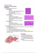
1.
Skeletal
Muscle
:
-
Striated
-
fibers/cells
are
large
in
diameter
and
also
length
-
Runs
the
entire
length
of
the
muscle
-
Multinucleated
cells
-
Controlled
by
the
somatic
nervous
system
2.
Cardiac
Muscle
:
-
Also
striated
-
Smaller
in
diameter
and
length
-
Specialized
junctions
between
the
cells
that
allow
the
cardiac
myocytes
to
contract
as
a
functional
unit
-
Controlled
by
the
autonomic
nervous
system
3.
Smooth
Muscle
:
-
Not
striated
-
Small,
single
nucleated
cells
-
Controlled
by
the
autonomic
nervous
system
-
Small
junctions
between
the
cells
for
cohesion
→
Shared
principles:
-
The
sliding
filament
mechanism
(myosin
filaments
bind
to
and
move
actin
filaments
to
shorten
the
muscle
cell)
-
Differences
in
organization
-
Myosin
and
actin
interactions
are
regulated
by
calcium
ions
-
Manner
will
be
different
between
the
three
muscle
types
-
Changes
in
the
membrane
potential
lead
to
contraction;
called
E-C
Coupling
(excitation
contraction
coupling)
Skeletal
Muscle:
-
Each
muscle
fiber
has
many
myofibrils,
which
number
determines
the
force
generating
capability
of
the
fiber.
-
Myofibrils
are
composed
of
many
sacomeres
that
are
in
series
(hundreds
of
thousands)
-
The
sarcomeres
are
what
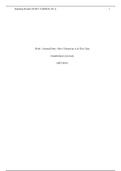
contract Sarcomere
Structure:
-
Alternating
light
and
dark
portions
(responsible
for
striations)
-
Z
lines
:
ends
of
the
sarcomeres
-
Thin
filaments
:
actin
-
Thick
filaments
:
myosin
(motor
properties)
-
A
Band:
the
dark
area
which
includes
the
entire
length
of
the
myosin
thick
filaments
-
I
Band:
the
light
area
where
there
is
only
actin
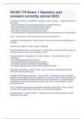
Sliding
Filament
Model
of
Contraction:
1.
Attachment
:
Myosin
heads
om
the
thick
filaments
attach
to
specific
sites
of
the
actin
thin
filaments,
forming
cross-bridges
2.
Power
Stroke
:
The
myosin
heads
pivot,
pulling
the
actin
filaments
toward
the
center
of
the
sarcomere.
→
this
action
is
powered
by
ATP;
when
ATP
binds
to
myosin
it
is
hydrolyzed
into
ADP
and
phosphate,
releasing
energy
that
allows
to
myosin
head
to
perform
a
power
stroke.
3.
Detachment
:
Another
ATP
molecule
binds
to
the
myosin
head,
causing
it
to
detach
from
the
actin
filament
4.
Recovery
:
The
myosin
head
returns
to
its
OG
position,
ready
to
form
another
cross-bridge
with
actin
5.
This
cycle
of
attachment,
power
stroke,
detachment,
and
recovery
repeats
numerous
times
during
a
muscle
contraction.
Each
cycle
pulls
the
actin
filaments
slightly
further
over
the
myosin
filaments,
which
shortens
the
entire
length
of
the
sarcomere
and
thus
the
muscle
fiber.
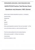
→
Regulation
by
Calcium:
-
Tropomyosin
:
runs
in
parallel
along
the
actin
and
binds
actin
at
the
site
where
myosin
would
bind
-
Prevents
myosin
from
binding
to
actin
when
there
is
no
calcium
present
-
Troponin
:
the
binding
of
calcium
to
troponin
causes
a
change
in
its
conformation
which
causes
tropomyosin
to
move,
exposing
the
actin
to
myosin
heads
4:
Muscle
Introduction
and
Contraction:
Muscle
Types:
→
Three
main
muscle
types
in
the
body…
1.
Skeletal
Muscle
:
-
Striated
-
fibers/cells
are
large
in
diameter
and
also
length
-
Runs
the
entire
length
of
the
muscle
-
Multinucleated
cells
-
Controlled
by
the
somatic
nervous
system
2.
Cardiac
Muscle
:
-
Also
striated
-
Smaller
in
diameter
and
length
-
Specialized
junctions
between
the
cells
that
allow
the
cardiac
myocytes
to
contract
as
a
functional
unit
-
Controlled
by
the
autonomic
nervous
system
3.
Smooth
Muscle
:
-
Not
striated
-
Small,
single
nucleated
cells
-
Controlled
by
the
autonomic
nervous
system
-
Small
junctions
between
the
cells
for
cohesion
→
Shared
principles:
-
The
sliding
filament
mechanism
(myosin
filaments
bind
to
and
move
actin
filaments
to
shorten
the
muscle
cell)
-
Differences
in
organization
-
Myosin
and
actin
interactions
are
regulated
by
calcium
ions
-
Manner
will
be
different
between
the
three
muscle
types
-
Changes
in
the
membrane
potential
lead
to
contraction;
called
E-C
Coupling
(excitation
contraction
coupling)
Skeletal
Muscle:
-
Each
muscle
fiber
has
many
myofibrils,
which
number
determines
the
force
generating
capability
of
the
fiber.
-
Myofibrils
are
composed
of
many
sacomeres
that
are
in
series
(hundreds
of
thousands)
-
The
sarcomeres
are
what
contract Sarcomere
Structure:
-
Alternating
light
and
dark
portions
(responsible
for
striations)
-
Z
lines
:
ends
of
the
sarcomeres
-
Thin
filaments
:
actin
-
Thick
filaments
:
myosin
(motor
properties)
-
A
Band:
the
dark
area
which
includes
the
entire
length
of
the
myosin
thick
filaments
-
I
Band:
the
light
area
where
there
is
only
actin
Sliding
Filament
Model
of
Contraction:
1.
Attachment
:
Myosin
heads
om
the
thick
filaments
attach
to
specific
sites
of
the
actin
thin
filaments,
forming
cross-bridges
2.
Power
Stroke
:
The
myosin
heads
pivot,
pulling
the
actin
filaments
toward
the
center
of
the
sarcomere.
→
this
action
is
powered
by
ATP;
when
ATP
binds
to
myosin
it
is
hydrolyzed
into
ADP
and
phosphate,
releasing
energy
that
allows
to
myosin
head
to
perform
a
power
stroke.
3.
Detachment
:
Another
ATP
molecule
binds
to
the
myosin
head,
causing
it
to
detach
from
the
actin
filament
4.
Recovery
:
The
myosin
head
returns
to
its
OG
position,
ready
to
form
another
cross-bridge
with
actin
5.
This
cycle
of
attachment,
power
stroke,
detachment,
and
recovery
repeats
numerous
times
during
a
muscle
contraction.
Each
cycle
pulls
the
actin
filaments
slightly
further
over
the
myosin
filaments,
which
shortens
the
entire
length
of
the
sarcomere
and
thus
the
muscle
fiber.
→
Regulation
by
Calcium:
-
Tropomyosin
:
runs
in
parallel
along
the
actin
and
binds
actin
at
the
site
where
myosin
would
bind
-
Prevents
myosin
from
binding
to
actin
when
there
is
no
calcium
present
-
Troponin
:
the
binding
of
calcium
to
troponin
causes
a
change
in
its
conformation
which
causes
tropomyosin
to
move,
exposing
the
actin
to
myosin
heads