Ways of Investigating the Brain
fMRI (functional magnetic resonance imaging)
Post Mortem
EEG (electroencephalograms)
ERP (event related potentials)
ERP
fMRI Post Mortem EEG >Application/ extension of EEG
>patient gets a task >analysis of brain after death >measures electrical activity through >Uses a stimulus (sensory/ cognitive/
>measures changes in blood flow >more likely used on those who had rare electrodes attached to scalp (skull cap) motor) to trigger brain waves
oxygenation as a result of brain activity disorders/ experienced unusual deficits >recordings represent brain wave patterns >Focuses on brain activity related to
>uses radio waves & magnetic field >damaged areas are examined to attempt generated from neurons stimuli/ areas of interest
>3D images, used to see what brain areas to establish likely cause of disorder >records general/overall activity linked to
are used to perform certain tasks different states (asleep/ aroused) + excellent temporal resolution
+ was vital for early understanding of key + can link ERP to exact cognitive functio
+ doesn’t rely on use of radiation brain processes + safe, non invasive
+ risk free, non invasive + improves medical knowledge + can identify activity in various brain - lack of standardisation methodology in
+ straight forward + detailed spatial resolution regions studies
+ high spatial resolution + good temporal resolution - poor spatial resolution
- ethical issues of consent - must be no background noise/ extrane
- expensive - issue of causation (observed damage may - very generalised nature of information variables to ensure data is pure
- poor temporal resolution, 5 second lag not be linked to the deficits, but to other - poor spatial resolution
- doesn’t measure individal neuron activity trauma/decay)
Functional Recovery
-example of neural plasticity
Biopsychology- Brain SS2
-following an injury, unaffected areas of brain are able to adapt & compensate for
damaged areas
-occurs quickly after trauma and then slows down Localisation of Function
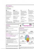
- lobes of the brain are associated with a specific function
During recovery, the brain rewires & reorganises itself, forming new synaptic - within each lobe are areas with certain functions
connections. - hemispheres control the opposite side of the body 2- Somatosensory Area
This involves a number of changes: 1- Motor Area
Front of parietal lobe (both hemispheres)
produces sensations in response to pleasure/ pain
Back of frontal lobe (both hemispheres)
different parts of the cortex are devoted to different parts of
> Axonal sprouting (growth of new nerve endings which connect with other undamaged responsible for voluntary movement on opposite side of body
controls what we sense (touch/ feel)
regions correspond to a certain area of body
nerve cells to form new neuronal pathways) injury= loss of coordination/ ability to perform fine motor movements
injury= vulnerable to numbness/ tingling sensations
>Reformation of blood vessels (because these may become severed from trauma) e
Lob
>Recruitment of homologous (similar) areas on opposite hemisphere Fro
ntal
6- Broca’s Area Par
>Secondary neural pathways are activated (dormant synapses) Left frontal lobe
ieta
l Lo
be
responsible for speech production
identified by Paul Broca after a post mortem 3- Visual Area
be
Case Study: Gabby Giffords of his patient Tan ital Lo
Occip Occipital lobe (both hemispheres)
damage= Broca’s aphasia- speech is slow, receives & processes visual information
shot in the head on January 8th 2011 (single bullet hit back of her head by only laborious & lacks fluency cross wired- info from right visual field is p
Tempo
crossing left hemisphere- bullet exited 9mm above left eyebrow) ral Lobe hemisphere
injury= chronic blindness, visual impairmen
initially placed in a coma, allow her brain to rest
5- Auditory Area
was able to respond to simple commands when periodically awakened Temporal lobe (both hemispheres)
analyses & processes speech/ acoustic based information (volume, tempo, 4- Wernicke’s Area
mid January- began simple physical therapy pitch)
Left temporal lobe- further back
responsible for language comprehension
her brain would need to recruit homologous areas on opposite hemisphere cross wired- info from left ear goes to right hemisphere
cross wired- info from right visual field is processed in left he
injury= inability to detect changes in pitch, difficulty understanding
damage= Wernicke’s aphasia- produces fluent but meaningle
Evaluation speech
(nonsense words= neologism)
P+ research support, E Gabby Giffords, recruitment of homologous areas on opposite Evaluation
hemisphere E suggests brain is able to adapt and compensate for damaged areas
P+ research support, E Petersen et al used brain scans to demonstrate how Wernicke and Broca’s area
P- problems with research support, E Gabby’s case was unique, only one study E active during different tasks, E suggests different parts of the brain have different functions, increase
of LoF
fMRI (functional magnetic resonance imaging)
Post Mortem
EEG (electroencephalograms)
ERP (event related potentials)
ERP
fMRI Post Mortem EEG >Application/ extension of EEG
>patient gets a task >analysis of brain after death >measures electrical activity through >Uses a stimulus (sensory/ cognitive/
>measures changes in blood flow >more likely used on those who had rare electrodes attached to scalp (skull cap) motor) to trigger brain waves
oxygenation as a result of brain activity disorders/ experienced unusual deficits >recordings represent brain wave patterns >Focuses on brain activity related to
>uses radio waves & magnetic field >damaged areas are examined to attempt generated from neurons stimuli/ areas of interest
>3D images, used to see what brain areas to establish likely cause of disorder >records general/overall activity linked to
are used to perform certain tasks different states (asleep/ aroused) + excellent temporal resolution
+ was vital for early understanding of key + can link ERP to exact cognitive functio
+ doesn’t rely on use of radiation brain processes + safe, non invasive
+ risk free, non invasive + improves medical knowledge + can identify activity in various brain - lack of standardisation methodology in
+ straight forward + detailed spatial resolution regions studies
+ high spatial resolution + good temporal resolution - poor spatial resolution
- ethical issues of consent - must be no background noise/ extrane
- expensive - issue of causation (observed damage may - very generalised nature of information variables to ensure data is pure
- poor temporal resolution, 5 second lag not be linked to the deficits, but to other - poor spatial resolution
- doesn’t measure individal neuron activity trauma/decay)
Functional Recovery
-example of neural plasticity
Biopsychology- Brain SS2
-following an injury, unaffected areas of brain are able to adapt & compensate for
damaged areas
-occurs quickly after trauma and then slows down Localisation of Function
- lobes of the brain are associated with a specific function
During recovery, the brain rewires & reorganises itself, forming new synaptic - within each lobe are areas with certain functions
connections. - hemispheres control the opposite side of the body 2- Somatosensory Area
This involves a number of changes: 1- Motor Area
Front of parietal lobe (both hemispheres)
produces sensations in response to pleasure/ pain
Back of frontal lobe (both hemispheres)
different parts of the cortex are devoted to different parts of
> Axonal sprouting (growth of new nerve endings which connect with other undamaged responsible for voluntary movement on opposite side of body
controls what we sense (touch/ feel)
regions correspond to a certain area of body
nerve cells to form new neuronal pathways) injury= loss of coordination/ ability to perform fine motor movements
injury= vulnerable to numbness/ tingling sensations
>Reformation of blood vessels (because these may become severed from trauma) e
Lob
>Recruitment of homologous (similar) areas on opposite hemisphere Fro
ntal
6- Broca’s Area Par
>Secondary neural pathways are activated (dormant synapses) Left frontal lobe
ieta
l Lo
be
responsible for speech production
identified by Paul Broca after a post mortem 3- Visual Area
be
Case Study: Gabby Giffords of his patient Tan ital Lo
Occip Occipital lobe (both hemispheres)
damage= Broca’s aphasia- speech is slow, receives & processes visual information
shot in the head on January 8th 2011 (single bullet hit back of her head by only laborious & lacks fluency cross wired- info from right visual field is p
Tempo
crossing left hemisphere- bullet exited 9mm above left eyebrow) ral Lobe hemisphere
injury= chronic blindness, visual impairmen
initially placed in a coma, allow her brain to rest
5- Auditory Area
was able to respond to simple commands when periodically awakened Temporal lobe (both hemispheres)
analyses & processes speech/ acoustic based information (volume, tempo, 4- Wernicke’s Area
mid January- began simple physical therapy pitch)
Left temporal lobe- further back
responsible for language comprehension
her brain would need to recruit homologous areas on opposite hemisphere cross wired- info from left ear goes to right hemisphere
cross wired- info from right visual field is processed in left he
injury= inability to detect changes in pitch, difficulty understanding
damage= Wernicke’s aphasia- produces fluent but meaningle
Evaluation speech
(nonsense words= neologism)
P+ research support, E Gabby Giffords, recruitment of homologous areas on opposite Evaluation
hemisphere E suggests brain is able to adapt and compensate for damaged areas
P+ research support, E Petersen et al used brain scans to demonstrate how Wernicke and Broca’s area
P- problems with research support, E Gabby’s case was unique, only one study E active during different tasks, E suggests different parts of the brain have different functions, increase
of LoF