Murmurs & ECG
interpretation
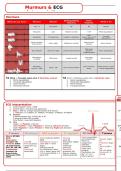
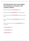
Murmurs
Distinguishing Extra
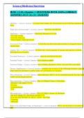
What do you hear? Murmur Where? What is it?
features features
HS I + II Everywhere Nil Nil Normal
Pansystolic Apex Radiates to axilla + AF? Mitral regurgitation
PSM
Tricuspid
Pansystolic Tricuspid area Loudest on inspiration Giant V wave in JVP
regurgitation
Pulsatile liver
Ejection Systolic Narrow pulse
Aortic region Radiates to carotids Aortic stenosis
(Cres-decres) pressure
Wide pulse
Early Diastolic Left sternal edge Loudest sitting forward pressure Aortic regurgitation
Collapsing pulse
Mid-diastolic Long angry
Mitral area Loudest w/ bell Mitral stenosis
rumbling murmur
Pansystolic As for murmurs Mixed murmur- mitral
Apex + LSE As for murmurs above
murmur + EDM above + aortic regurgitation
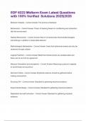
1st 2nd 1st
S1 Mitral + Tricuspid valves shut Ventricles contract S2 Aortic + Pulmonary valves shut Ventricles relax
1. Mitral regurgitation 1. Aortic regurgitation
2. Tricuspid regurgitation 2. Pulmonary regurgitation
3. Aortic stenosis 3. Mitral stenosis
4. Pulmonary stenosis 4. Tricuspid stenosis
ECG
ECG
Ab
Abn
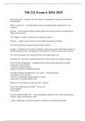
ECG interpretation
1. Confirm patient name + DOB
2. Confirm date + time ECG was performed
3. Rate = 300/ no. of large squares between R-R interval QRS complex 1st
1 =300 bpm, 2= 150bpm, 3= 100bpm, 4=75bpm, 5=60bpm, 6=50bpm
4. Rhythm
P waves present? Yes Sinus; No AF tach
P waves precede QRS complex?
Atrial flutter = saw-tooth baseline
5. Axis
Leads I + II Positive = Normal T wave
Lead I Positive + Lead II Negative = ‘Leaving’ Left-axis deviation P wave 2nd
Lead I Negative + Lead II Positive = ‘Reaching’ Right-axis deviation ST segment
M
6. Segments I/W
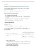
P waves PR interval QRS complex ST segment PR
QT interval
interval QT interval
T waves
Absent AF/SAN Atri
Toxins (macrolides,
block Wide conduction defect/ Elevated
anti-arrhythmias, Peaked: hyper-
Dissociated WPW Acute MI
TCAs, H2 K+
Complete HB Long HB Pathological Q wave > Pericarditis: saddle-
antagonists) Flattened: hypo-
P-mitrale (bifid) Short accessory 1mm wide +>2mm shaped
Inherited K+
LA hypertrophy, conduction e.g. depth Ventricular
Myocarditis Normal aVR + Mo
HTN, AS, MR, MS WPW Full thickness MI aneurysm
Mitral valve V1
P-pulmonale Depressed RVH dominant R wave Depressed
prolapse V2 +V3 (Afro)
(peaked) RA pericarditis (V1) + deep S wave (V6) Ischaemia: flat
Electrolytes: low Abnormal I, II,
hypertrophy, LVH R wave (V5/V6) + S Digoxin: sloping
Mg2+, K+, Ca2+, V4-V6
pulmonary HTN, wave (V1) down/reverse tick
temp (J waves) fib
COPD
interpretation
Murmurs
Distinguishing Extra
What do you hear? Murmur Where? What is it?
features features
HS I + II Everywhere Nil Nil Normal
Pansystolic Apex Radiates to axilla + AF? Mitral regurgitation
PSM
Tricuspid
Pansystolic Tricuspid area Loudest on inspiration Giant V wave in JVP
regurgitation
Pulsatile liver
Ejection Systolic Narrow pulse
Aortic region Radiates to carotids Aortic stenosis
(Cres-decres) pressure
Wide pulse
Early Diastolic Left sternal edge Loudest sitting forward pressure Aortic regurgitation
Collapsing pulse
Mid-diastolic Long angry
Mitral area Loudest w/ bell Mitral stenosis
rumbling murmur
Pansystolic As for murmurs Mixed murmur- mitral
Apex + LSE As for murmurs above
murmur + EDM above + aortic regurgitation
1st 2nd 1st
S1 Mitral + Tricuspid valves shut Ventricles contract S2 Aortic + Pulmonary valves shut Ventricles relax
1. Mitral regurgitation 1. Aortic regurgitation
2. Tricuspid regurgitation 2. Pulmonary regurgitation
3. Aortic stenosis 3. Mitral stenosis
4. Pulmonary stenosis 4. Tricuspid stenosis
ECG
ECG
Ab
Abn
ECG interpretation
1. Confirm patient name + DOB
2. Confirm date + time ECG was performed
3. Rate = 300/ no. of large squares between R-R interval QRS complex 1st
1 =300 bpm, 2= 150bpm, 3= 100bpm, 4=75bpm, 5=60bpm, 6=50bpm
4. Rhythm
P waves present? Yes Sinus; No AF tach
P waves precede QRS complex?
Atrial flutter = saw-tooth baseline
5. Axis
Leads I + II Positive = Normal T wave
Lead I Positive + Lead II Negative = ‘Leaving’ Left-axis deviation P wave 2nd
Lead I Negative + Lead II Positive = ‘Reaching’ Right-axis deviation ST segment
M
6. Segments I/W
P waves PR interval QRS complex ST segment PR
QT interval
interval QT interval
T waves
Absent AF/SAN Atri
Toxins (macrolides,
block Wide conduction defect/ Elevated
anti-arrhythmias, Peaked: hyper-
Dissociated WPW Acute MI
TCAs, H2 K+
Complete HB Long HB Pathological Q wave > Pericarditis: saddle-
antagonists) Flattened: hypo-
P-mitrale (bifid) Short accessory 1mm wide +>2mm shaped
Inherited K+
LA hypertrophy, conduction e.g. depth Ventricular
Myocarditis Normal aVR + Mo
HTN, AS, MR, MS WPW Full thickness MI aneurysm
Mitral valve V1
P-pulmonale Depressed RVH dominant R wave Depressed
prolapse V2 +V3 (Afro)
(peaked) RA pericarditis (V1) + deep S wave (V6) Ischaemia: flat
Electrolytes: low Abnormal I, II,
hypertrophy, LVH R wave (V5/V6) + S Digoxin: sloping
Mg2+, K+, Ca2+, V4-V6
pulmonary HTN, wave (V1) down/reverse tick
temp (J waves) fib
COPD