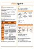
Gallstones & Jaundice
Gallstones
24% of women and 12% of men may develop local infection + cholecystitis
Jaundice
Definition jaundice, also known as icterus I the yellow discolouration of mucous
Type of gallstones membrane, sclera + skin due to the accumulation of bilirubin
Cholesterol – light yellow to dark green, usually solitary, with a dark, central spot. Need to Jaundice may be seen @ a [>2.5-3.0mg/dl/42.8-51.3mmol/l]
be > 80% cholesterol by weight Aetiology
Pigment- also known as bilirubin stones, are dark/black and numerous. Predominately Pre-hepatic Intrahepatic Post-hepatic
composed of bilirubin and calcium salts that are found in bile. Contain < 20% cholesterol Crigler-Najjar syndrome Viral + drug induced
Mixed usually brown in colour, contain 20-80% cholesterol, also made up of calcium Gilbert’ syndrome hepatitis Gallstones in bile duct
carbonate and other bile pigments Haemolysis e.g. Alcohol liver disease Pancreatic cancer
Gallstones + bile duct obstruction cholangitis (due to stagnate bile causing mucosal thalassaemia, sickle cell Hepatic cirrhosis Schistosomiasis
irritation) disease Primary biliary cirrhosis Biliary atresia
Gallstones + Ampulla of Vater obstruction pancreatitis Drugs e.g. Rifampicin Leptospirosis Cholangiocarcinoma
RF Four F’s Female Fat (obesity) Fertile (pregnancy) Forty Malaria Physiological neonatal Mirizzi’s syndrome
Haemolytic uraemic jaundice
Signs + symptoms syndrome
Colicky RUQ pain that occurs post-prandially
Ix
Worse following a fatty meal when cholecystokinin levels are highest + gallbladder
Establish the type of jaundice
contraction is maximal
1. Appearance of urine + stools?
Gallstone- 2. LFTs?
related Signs + symptoms Management 3. Bilirubin levels?
disease 4. Alkaline phosphatase levels?
Colicky abdominal pain, If imaging + history confirmative
Biliary colic worse post-prandially + Laparoscopic cholecystectomy Pre-hepatic Intrahepatic Post-hepatic
after fatty foods Urine Normal Dark Dark
Acute Imaging USSS + cholecystectomy (w/i Stool Normal Pale Pale
RUQ pain, fever, Murphy’s
cholecystiti 48hrs) Conjugated
sign, mildly deranged LFTs Normal High High
s bilirubin
Imaging w/ USS +/- CT ideally, surgery Unconjugated
bilirubin
Normal/Raised High Normal
Prodromal illness + RUQ although subtotal cholecystectomy may be
Gallbladder
abscess
pain, swinging fever + needed if Calot’s triangle is hostile (space Total bilirubin Normal/Raised High High
systemically unwell between common hepatic duct, cystic duct Alkaline
and inferior hepatic border) phosphatase
Normal High Very high
Fluid resuscitation
Severely septic patient USS of liver + biliary tree
Broad-spectrum IV Abx
Cholangitis Jaundice Identify gallstones? Pancreatic masses? bile duct calibre?
Correct any coagulopathy
RUQ pain If pancreatic neoplasia suspected CT scan
Early ERCP
Laparotomy + removal of the gallstone Liver tumours/Cholangiocarcinoma MRI/MRCP MRCP fails? ERCP
Hx of cholangitis Enterotomy must be made proximal to the
Gallstone Mx
Known gallstones site of obstruction + not @ the site. If a
ileus Malignancy tent inserted (metal/plastic)/failed? drainage of biliary system
Small bowel obstruction fistula between duodenum + gallbladder is
present don’t interfere percutaneously via a transhepatic route
Bile duct injury surgery
Risks of ERCP Gallstones removed by ERCP + cholecystectomy performed
Bleeding (0.9%), duodenal perforation (0.4%), cholangitis (1.1%), pancreatitis (1.5%) Cholangitis high dose broad-spectrum Abx via IV route + biliary decompression