Summary Muscular, Nervous and
Skeletal – minor PT
Book Chapter 1
The Muscular System
Muscles generate force when they are activated. This is referred to as a muscle
contraction or muscle action. Of the three types of muscle:
Smooth
Cardiac
Skeletal
The Skeletal type attaches to bones, causing them to rotate around joints. It is this
function of skeletal muscles that allows us to run, jump, and lift and throw things. The
function of muscle is dictated by its structure.
Gross Anatomy of Skeletal Muscle
See book page 4. Figure 1.1 > or anatomy summary
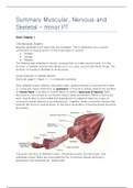
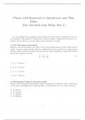
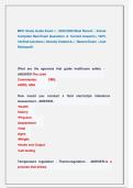
Each skeletal muscle (deltoid, pectoralis major, gastrocnemius) is surround by a layer
of connective tissue referred to as epimysium. A muscle is further divided into bundles
of muscle fibers. A bundle of muscle fibers is called a fasciculus of fascicle. Each
fasciculus is surrounded by connective tissue called perimysium. Within a fasciculus,
each muscle fiber is surrounded and separated from adjacent fibers by a layer of
connective tissue referred to as endomysium. Together, these connective tissues help
transmit the force of muscle action to the bone via another connective tissue structure,
the tendon.
The gross structure of skeletal muscle. The whole muscle, the fasciculus, and
individual muscle fibers are surrounded by the connective tissues epimysium,
perimysium and endomysium, respectively.
,Microscopic Anatomy of Skeletal Muscle
Each muscle fiber is a cell, with many of the same structural components as other
cells. Each muscle fiber is surrounded by a plasma membrane, referred to as the
sarcolemma. The sarcolemma encloses the contents of the cell, regulates the passage
of materials such as glucose into and out of the cell, and receives and conducts stimuli
in the form of electrical impulses of action potentials.
Skeletal muscle cells are multinucleated, so they possess more than one nucleus. The
nuclei contain the genetic material, or DNA, of the cell, and are largely responsible for
initiating the processes associated with adaptations to exercise, such as muscle cell
enlargement or hypertrophy.
Adaptations to resistance training and aerobic endurance training are discussed in
Chapter 5 and 6.
Within the sarcolemma and outside the nuclei there is the cytoplasm, and called
sarcoplasm in muscle. This watery solution contains the cell’s energy sources, such as
Adenosine triphosphate (ATP).
Phosphocreatine
Glycogen
Fat droplets.
ATP = the only direct source of energy for muscle action.
Also suspended within the sarcoplasm are organelles.
These include mitochondria (singular is mitochondrion), which are the sites of
aerobic ATP production within the cell and thus of great importance for aerobic
exercise performance.
Another important organelle is the sarcoplasmic reticulum. This organelle stores
calcium and regulate the muscle action process by altering the intracellular
calcium concentration. Specifically, the sarcoplasmic reticulum releases calcium
into the sarcoplasm of the cell when an action potential passes to the interior of
the cell via structures called transverse tubules of T-tubules.
o The T-tubules are channels that form from openings in the sarcolemma
of the muscle cell.
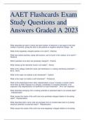
Myofibril
Structures that run parallel to the length of the muscle fiber are known as myofibrils.
Each myofibril is a bundle of myofilaments, which primarily consist of myosin (thick
filaments) and actin (thin filaments). The movement of myosin over actin giving
contraction in muscles. Because each myosin consist of a head (and neck and tail)
which is capable of attaching to and pulling on the actin filament. The energy from
hydrolysis (=splitting) of ATP is used to perform this power stroke, an important step in
the process of muscle activation.
Each actin filament is formed form G-actin proteins (individual globular). Each G-actin
has a binding site for a myosin head. The G-actin proteins assemble into strands of F-
actin (filamentous). Associated with the actin filament are two other protein structures:
Tropomyosin (a rod-like protein that spans the length of seven G-actin proteins
along the length of the actin filament).
, Troponin (each tropomyosin is attaches – at the end – to troponin, when bound
to calcium, troponin causes the movement of tropomyosin away from the
myosin binding sites on actin).
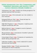
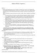
Sarcomere
The basic functional and contractile unit of skeletal muscle is the sarcomere.
Z-line: Sarcomere extends from one Z-line to an adjacent Z-line. Actin filaments
are anchored at one end of the Z-line.
M-line: In the middle of the H-zone. The M-line helps align adjacent myosin
filaments.
A-band: The A-band is determined by the width of a myosin filament. It is the A-
band that provides the dark striation of skeletal muscle.
I-band: Spans the distance between the ends of adjacent myosin filaments. Each
I-band lies partly in each of two sarcomeres.
H-zone: The area of the A-band that contains myosin, but nog actin, is the H-
zone.
Sarcomere: The contraction of an sarcomere works within 6 steps.
Skeletal – minor PT
Book Chapter 1
The Muscular System
Muscles generate force when they are activated. This is referred to as a muscle
contraction or muscle action. Of the three types of muscle:
Smooth
Cardiac
Skeletal
The Skeletal type attaches to bones, causing them to rotate around joints. It is this
function of skeletal muscles that allows us to run, jump, and lift and throw things. The
function of muscle is dictated by its structure.
Gross Anatomy of Skeletal Muscle
See book page 4. Figure 1.1 > or anatomy summary
Each skeletal muscle (deltoid, pectoralis major, gastrocnemius) is surround by a layer
of connective tissue referred to as epimysium. A muscle is further divided into bundles
of muscle fibers. A bundle of muscle fibers is called a fasciculus of fascicle. Each
fasciculus is surrounded by connective tissue called perimysium. Within a fasciculus,
each muscle fiber is surrounded and separated from adjacent fibers by a layer of
connective tissue referred to as endomysium. Together, these connective tissues help
transmit the force of muscle action to the bone via another connective tissue structure,
the tendon.
The gross structure of skeletal muscle. The whole muscle, the fasciculus, and
individual muscle fibers are surrounded by the connective tissues epimysium,
perimysium and endomysium, respectively.
,Microscopic Anatomy of Skeletal Muscle
Each muscle fiber is a cell, with many of the same structural components as other
cells. Each muscle fiber is surrounded by a plasma membrane, referred to as the
sarcolemma. The sarcolemma encloses the contents of the cell, regulates the passage
of materials such as glucose into and out of the cell, and receives and conducts stimuli
in the form of electrical impulses of action potentials.
Skeletal muscle cells are multinucleated, so they possess more than one nucleus. The
nuclei contain the genetic material, or DNA, of the cell, and are largely responsible for
initiating the processes associated with adaptations to exercise, such as muscle cell
enlargement or hypertrophy.
Adaptations to resistance training and aerobic endurance training are discussed in
Chapter 5 and 6.
Within the sarcolemma and outside the nuclei there is the cytoplasm, and called
sarcoplasm in muscle. This watery solution contains the cell’s energy sources, such as
Adenosine triphosphate (ATP).
Phosphocreatine
Glycogen
Fat droplets.
ATP = the only direct source of energy for muscle action.
Also suspended within the sarcoplasm are organelles.
These include mitochondria (singular is mitochondrion), which are the sites of
aerobic ATP production within the cell and thus of great importance for aerobic
exercise performance.
Another important organelle is the sarcoplasmic reticulum. This organelle stores
calcium and regulate the muscle action process by altering the intracellular
calcium concentration. Specifically, the sarcoplasmic reticulum releases calcium
into the sarcoplasm of the cell when an action potential passes to the interior of
the cell via structures called transverse tubules of T-tubules.
o The T-tubules are channels that form from openings in the sarcolemma
of the muscle cell.
Myofibril
Structures that run parallel to the length of the muscle fiber are known as myofibrils.
Each myofibril is a bundle of myofilaments, which primarily consist of myosin (thick
filaments) and actin (thin filaments). The movement of myosin over actin giving
contraction in muscles. Because each myosin consist of a head (and neck and tail)
which is capable of attaching to and pulling on the actin filament. The energy from
hydrolysis (=splitting) of ATP is used to perform this power stroke, an important step in
the process of muscle activation.
Each actin filament is formed form G-actin proteins (individual globular). Each G-actin
has a binding site for a myosin head. The G-actin proteins assemble into strands of F-
actin (filamentous). Associated with the actin filament are two other protein structures:
Tropomyosin (a rod-like protein that spans the length of seven G-actin proteins
along the length of the actin filament).
, Troponin (each tropomyosin is attaches – at the end – to troponin, when bound
to calcium, troponin causes the movement of tropomyosin away from the
myosin binding sites on actin).
Sarcomere
The basic functional and contractile unit of skeletal muscle is the sarcomere.
Z-line: Sarcomere extends from one Z-line to an adjacent Z-line. Actin filaments
are anchored at one end of the Z-line.
M-line: In the middle of the H-zone. The M-line helps align adjacent myosin
filaments.
A-band: The A-band is determined by the width of a myosin filament. It is the A-
band that provides the dark striation of skeletal muscle.
I-band: Spans the distance between the ends of adjacent myosin filaments. Each
I-band lies partly in each of two sarcomeres.
H-zone: The area of the A-band that contains myosin, but nog actin, is the H-
zone.
Sarcomere: The contraction of an sarcomere works within 6 steps.