HHIS221: HUMAN HISTOLOGY
Lesson 6 | Muscle Tissue
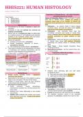
Organization of Skeletal Muscle – also called Interstitial
connective tissue of our muscle
Topic Outline:
● Muscle Tissue Layers of Connective Tissue present in all types of muscle;
● Skeletal Muscle seen well in skeletal muscle (collagen are present in
● Cardiac Muscle connective tissue layer of our muscles, to transmit the
● Smooth Muscle mechanical forces generated by our muscle cell for our muscle
Muscle Tissue fiber)
● Characterized by the ability to contract or move upon 1) Epimysium – an external sheath of dense irregular
stimulation. connective tissue, surrounds the entire muscle (carries
● Composed of cells that optimize the universal cell large nerves, blood vessels, and lymphatics)
property of contractility. 2) Perimysium - thin connective tissue layer that
● Muscle cells are of mesodermal origin and differentiate immediately surrounds each bundle of muscle fibers
by a gradual process of cell lengthening with abundant termed a fascicle (nerves, blood vessels, and lymphatics
synthesis of the myofibrillar proteins actin and myosin. penetrate perimysium to supply nutrients to each
(Two contractile proteins – actin and myosin) fascicles)
● Muscle cell organelles 3) Endomysium – within each fascicle is a thin delicate layer
➔ Cytoplasm of muscle cells - sarcoplasm of reticular fibers tissue (with scattered fibroblasts)
➔ Smooth Endoplasmic Reticulum of Muscle – surrounding the external lamina of individual muscle
sarcoplasmic reticulum fibers.
➔ Cell membrane and external lamina - sarcolemma 4) Deep Fascia – Dense Irregular Connective Tissue
● Three types of muscles can be distinguished on the basis overlying epimysium
of morphologic and functional characteristics with the 5) Myotendinous junctions – join the muscle to bone, skin,
structure of each adapted to its physiological role. or another muscle
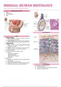
Organization Within Muscle Fibers
● Tissues are sectioned in three ways:
● Striations show alternating light and dark bands
Skeletal Muscle (Longitudinal action):
● Striated muscle, Voluntary muscle – appearance ➔ Dark bands are called A Bands (Anisotropic)
prominent cross striations ➔ Light bands are called I Bands (Isotropic)
● Responsible for the movement of the skeleton as well as
organs such as the globe of the eye and the tongue
● consists of muscle fibers, which are long, cylindrical
multinucleated cells with diameters of 10-100 μm
● Development of skeletal muscle
1) During embryonic muscle development,
mesenchymal myoblasts fuse, forming myotubes with
many nuclei. ● Sarcoplasm is highly organized, containing primarily long
2) Myotubes then further differentiate to form striated cylindrical filament bundles called myofibrils
muscle fibers. ● I bands are bisected by a dark transverse line, the Z disc.
3) Satellite cells proliferate and produce new muscle ● The repetitive functional subunit of the contractile
fibers following muscle injury. – located at the external apparatus, the sarcomere, extends from Z disc to Z disc
surface of muscle fiber
1
Lesson 6 | Muscle Tissue
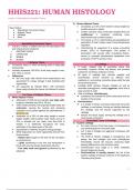
Organization of Skeletal Muscle – also called Interstitial
connective tissue of our muscle
Topic Outline:
● Muscle Tissue Layers of Connective Tissue present in all types of muscle;
● Skeletal Muscle seen well in skeletal muscle (collagen are present in
● Cardiac Muscle connective tissue layer of our muscles, to transmit the
● Smooth Muscle mechanical forces generated by our muscle cell for our muscle
Muscle Tissue fiber)
● Characterized by the ability to contract or move upon 1) Epimysium – an external sheath of dense irregular
stimulation. connective tissue, surrounds the entire muscle (carries
● Composed of cells that optimize the universal cell large nerves, blood vessels, and lymphatics)
property of contractility. 2) Perimysium - thin connective tissue layer that
● Muscle cells are of mesodermal origin and differentiate immediately surrounds each bundle of muscle fibers
by a gradual process of cell lengthening with abundant termed a fascicle (nerves, blood vessels, and lymphatics
synthesis of the myofibrillar proteins actin and myosin. penetrate perimysium to supply nutrients to each
(Two contractile proteins – actin and myosin) fascicles)
● Muscle cell organelles 3) Endomysium – within each fascicle is a thin delicate layer
➔ Cytoplasm of muscle cells - sarcoplasm of reticular fibers tissue (with scattered fibroblasts)
➔ Smooth Endoplasmic Reticulum of Muscle – surrounding the external lamina of individual muscle
sarcoplasmic reticulum fibers.
➔ Cell membrane and external lamina - sarcolemma 4) Deep Fascia – Dense Irregular Connective Tissue
● Three types of muscles can be distinguished on the basis overlying epimysium
of morphologic and functional characteristics with the 5) Myotendinous junctions – join the muscle to bone, skin,
structure of each adapted to its physiological role. or another muscle
Organization Within Muscle Fibers
● Tissues are sectioned in three ways:
● Striations show alternating light and dark bands
Skeletal Muscle (Longitudinal action):
● Striated muscle, Voluntary muscle – appearance ➔ Dark bands are called A Bands (Anisotropic)
prominent cross striations ➔ Light bands are called I Bands (Isotropic)
● Responsible for the movement of the skeleton as well as
organs such as the globe of the eye and the tongue
● consists of muscle fibers, which are long, cylindrical
multinucleated cells with diameters of 10-100 μm
● Development of skeletal muscle
1) During embryonic muscle development,
mesenchymal myoblasts fuse, forming myotubes with
many nuclei. ● Sarcoplasm is highly organized, containing primarily long
2) Myotubes then further differentiate to form striated cylindrical filament bundles called myofibrils
muscle fibers. ● I bands are bisected by a dark transverse line, the Z disc.
3) Satellite cells proliferate and produce new muscle ● The repetitive functional subunit of the contractile
fibers following muscle injury. – located at the external apparatus, the sarcomere, extends from Z disc to Z disc
surface of muscle fiber
1