Experimentation II – Literature & Lecture Slides
General Introduction
Invasiveness Technique
Invasive -Single cell recordings -Deep brain stimulation
-Brain surgery -Pharmacology
-Electro-convulsive therapy
Somewhat invasive -PET -CT
-regional cerebral blood flow (rCBF) -rTMS
Non-invasive -Single pulse TMS -Peripheral recordings
-EEG -Saliva checks
-MEG -Neural network modeling
-Optical imaging -Behavioral genetics
-(f)MRI -Affect/Mood induction
-Eye tracking -Circadian rhythm
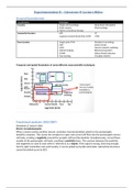
Temporal and Spatial Resolution of some different neuroscientific techniques
Functional analysis (EEG/ERP)
Woodman & Lecture slides
Electro-encephalography
When a neuron excites another neuron, excitatory neurotransmitters attach to the postsynaptic
dendritic receptors. This causes the receptors to open and current will flow into the postsynaptic neuron
cell body, creating a negativity around the synaptic cleft and the dendrite. Simultaneously, current flows
outside of the postsynaptic cell body, resulting in positivity there. This contrast between the positivity
and negativity so close to each other is referred to as a dipole. If this signal is strong, lasts long enough,
has the right orientation and synchronicity, it can be picked up by EEG electrodes. Subcortical structures
cannot be picked up on by EEG.
, The electrical activity in the brain is never
random. EEG mostly picks up on distinct rhythms
of oscillations (= rhythmic waves in electrical
current) which vary in frequency (slow-fast | 0-70
Hz), amplitude (low-high | -50 – 50 mV), phase
(in the sine cycle | 0-360 degrees) and timing.
Specific oscillations have been identified and
related to specific behaviors (e.g., alpha waves
are related to relaxation). EEG can identify the
location (topography) of the signal, although it is
not very spatially specific. You can also model the
source of the signal as a dipole.
The raw EEG signal is a sum of all the frequencies that are being picked up in that area. Fourier
transformation can split this sum back up into its part, allowing us to study the different frequencies
separately.
Different oscillations can show coherence or synchronization, which often reflects communication
between brain areas. Slower waves can carry faster waves to travel further along the brain.
When measuring EEG you always have to have reference electrodes which you can use to subtract noise
from your signal. Which reference place you choose can influence your results. You can place them
somewhere on the body (mastoids, nose, earlobes, shoulder) or you can take the average of all your
electrodes and subtract that.
The electric signal that is being picked up is referred to as a brain potential. Thus, an EEG session results
in a long time series of brain potentials on all the places the electrodes have been put. EEG signals that
occur systematically and synchronous to a meaningful event (e.g., presentation of a stimulus, or
moment of response) are called event-related potentials (ERPs).
ERPs are extremely small. All brain potentials have to be amplified before they can be digitized and
shown on a computer screen. However, because they are so small they are also very easily drowned by
noise. It is therefore very important that you take precautions to avoid noise and filter out noise during
analysis (prevention is better).
Maximize signal:
Minimize skin impedance by scratching the skin with a blunt needle
Maximize conductivity by using electrode gel
Acquire enough trials
Always check your set-up (broken electrodes)
Minimize noise:
Put electrodes around the eyes so you can correct for eye movement during analysis
Guard your experimenting room from power sources (e.g., elevators, new computers)
Minimize motion
Check that your electrodes are adequately connected
Make sure the cap is in the right location
Keep the experimenting room cool to avoid sweating
Cleaning EEG data
General Introduction
Invasiveness Technique
Invasive -Single cell recordings -Deep brain stimulation
-Brain surgery -Pharmacology
-Electro-convulsive therapy
Somewhat invasive -PET -CT
-regional cerebral blood flow (rCBF) -rTMS
Non-invasive -Single pulse TMS -Peripheral recordings
-EEG -Saliva checks
-MEG -Neural network modeling
-Optical imaging -Behavioral genetics
-(f)MRI -Affect/Mood induction
-Eye tracking -Circadian rhythm
Temporal and Spatial Resolution of some different neuroscientific techniques
Functional analysis (EEG/ERP)
Woodman & Lecture slides
Electro-encephalography
When a neuron excites another neuron, excitatory neurotransmitters attach to the postsynaptic
dendritic receptors. This causes the receptors to open and current will flow into the postsynaptic neuron
cell body, creating a negativity around the synaptic cleft and the dendrite. Simultaneously, current flows
outside of the postsynaptic cell body, resulting in positivity there. This contrast between the positivity
and negativity so close to each other is referred to as a dipole. If this signal is strong, lasts long enough,
has the right orientation and synchronicity, it can be picked up by EEG electrodes. Subcortical structures
cannot be picked up on by EEG.
, The electrical activity in the brain is never
random. EEG mostly picks up on distinct rhythms
of oscillations (= rhythmic waves in electrical
current) which vary in frequency (slow-fast | 0-70
Hz), amplitude (low-high | -50 – 50 mV), phase
(in the sine cycle | 0-360 degrees) and timing.
Specific oscillations have been identified and
related to specific behaviors (e.g., alpha waves
are related to relaxation). EEG can identify the
location (topography) of the signal, although it is
not very spatially specific. You can also model the
source of the signal as a dipole.
The raw EEG signal is a sum of all the frequencies that are being picked up in that area. Fourier
transformation can split this sum back up into its part, allowing us to study the different frequencies
separately.
Different oscillations can show coherence or synchronization, which often reflects communication
between brain areas. Slower waves can carry faster waves to travel further along the brain.
When measuring EEG you always have to have reference electrodes which you can use to subtract noise
from your signal. Which reference place you choose can influence your results. You can place them
somewhere on the body (mastoids, nose, earlobes, shoulder) or you can take the average of all your
electrodes and subtract that.
The electric signal that is being picked up is referred to as a brain potential. Thus, an EEG session results
in a long time series of brain potentials on all the places the electrodes have been put. EEG signals that
occur systematically and synchronous to a meaningful event (e.g., presentation of a stimulus, or
moment of response) are called event-related potentials (ERPs).
ERPs are extremely small. All brain potentials have to be amplified before they can be digitized and
shown on a computer screen. However, because they are so small they are also very easily drowned by
noise. It is therefore very important that you take precautions to avoid noise and filter out noise during
analysis (prevention is better).
Maximize signal:
Minimize skin impedance by scratching the skin with a blunt needle
Maximize conductivity by using electrode gel
Acquire enough trials
Always check your set-up (broken electrodes)
Minimize noise:
Put electrodes around the eyes so you can correct for eye movement during analysis
Guard your experimenting room from power sources (e.g., elevators, new computers)
Minimize motion
Check that your electrodes are adequately connected
Make sure the cap is in the right location
Keep the experimenting room cool to avoid sweating
Cleaning EEG data