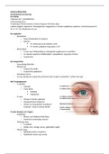
Samenvatting OOG
HC Anatomie van het oog
De Orbita:
• Bestaat uit 7 schedelbotten
• Peervormig 30 cc
• Opening is 45mm breed en 35mm hoog en 40-45mm diep
• Bevat oogbol, oogzenuw, traanapparaat, oogspieren,m. levator palpebrae superieur, hersenzenuwen (II,
III, IV, V1, VI), bloedvaten en vet
De oogleden:
- Buitenblad
o Huid, bindweefsel & wimpers
o Spieren
M. orbicularis oculi (sluiten, nVII)
M. levator palpabre (oog open, n.III)
- Binnenblad
o Tarsus met Meibomklieren (stevigheid ooglidrand en traanfilm)
o M. tarsalis superieur (Müllerspier): sympathisch, oog open, Horner
o Conjunctiva
De Conjunctiva:
- Doorzichtig slijmvlies
- Bestaat uit:
o Conjunctiva bulbi
o Conjunctiva palpebrae
- Overgang: fornix
- Let op: dankzij de conjunctiva (fornices) kan er geen contactlens “achter het oog”
Het Traanapparaat:
- Traanklier
o Lateraal
o Twee delen
Orbitaal
Palpabraal
- Tranen
o Uitvoer in fornix superieur
o Verspreid door knipperen
o Afvoer via traanpunten (mediaal) >
traanzak > ductus nasolacrimalis
Tranen bestaan uit 3 lagen:
- Lipidelaag
o Klieren van Meibom/Moll/Zeiss
o Voorkomt verdamping tranen
- Waterige laag
o Traanklier
o Helder zien, reinigt cornea, glijmiddel ooglid
- Mucine laag
o Slijmbekercellen conjuctiva
o Stabilisatie tranen op cornea
,De Functie van de oogspieren:
n. Oculomotorius (n.III):
- m. rectus medialis
- m. rectus superior
- m. rectus inferior
- m. obliquus inferior
- (m. levator palpebrae)
- (m. sphincter pupillae)
- (m. ciliaris)
n. Trochlearis (n.IV):
• m. obliquus superior
n. Abducens (n.VI):
• m. rectus lateralis
Kijken:
• Achter in het oog bevindt zich het
netvlies.
• Het netvlies vangt alle visuele
informatie in het oog en geeft het
door aan de oogzenuw.
• Hierna wordt alle visuele informatie in de hersenen verwerkt. Zo ervaren wij “zien’’
Macroanatomie: van voor naar achter
• Het voorsegment:
– De voorste oogkamer: tussen cornea en iris ≅ 3mm diep, 0.25 mL
– Achterste oogkamer: tussen iris en lens ≈ 0.06 mL
• Het achtersegment:
– Glasvochtholte ≈5-6 mL
• Totale volume ≈ 6-7 mL
• Lengte ≈24 mm
Macroanatomie: van buiten naar binnen
• Buitenste laag:
– Cornea
– Sclera (bedekt met conjunctiva, episclera en capsule van tenon)
– (Limbus)
• Middelste laag (onder de sclera)
– Uvea:
• Iris
• Corpus ciliare
• Choroid
• Binnenste laag (onder de sclera)
– Netvlies (retina)
De Sclera:
• Opaak en stevig (collageen): “exoskelet”
• Dunste achter de lateralis spieren (0.3mm)
• Dikste posterieur (1mm)
• Zeef bij oorsprong n.opticus (lamina cribrosa)
De Voorste Oogkamer (VOK)
• ≈3mm diep van cornea endotheel tot iris/pupil/lens
• Wordt met toenemende leeftijd ondieper
• Bevat kamerwater (voeding cornea en lens)
,• Kamerhoek 360 graden rondom
Flow Kamerwaterproductie
• Productie in corpus ciliare (2uL/min)
• Tussen de lens en de iris
• Door de pupil
• Naar de kamerhoek
• Trabekelsysteem > kanaal van Schlemm > watervenen
Iris:
• Scotopisch (donker)
– Mydriasis
– M. dilatator pupillae
• Mesoptisch
• Fotopisch (licht)
– Miosis
– M. Sfincter pupillae
De lens:
• Functie: accomodatie, UV filter, licht doorlaten, lichtstralen bundelen
• “opgehangen” door zonulavezels
• Met toenemend leeftijd minder accommodatie en helderheid (“staar”)
Posterieur Segment:
- Bevat glasvocht
o Glasvochtmembraan gevuld met gelei
o 98% water en 2% hyaluronzuur en collageen
o vervloeit bij ouder worden (waardoor risico op netvliesscheur en loslating!)
, De Fundus:
• Nervus opticus
• a. centralis retinae
• v. centralis retinae
• Retina: macula
De Retina:
• Begint voorbij de pars plana van corpus ciliare (Ora Serrata)
• Transparante binnenbekleding van het oog
• Vangt visuele informatie en geeft het door aan de oogzenuw
De Retina:
- Licht > Media, Ganglion cellaag, Bipolaire cellaag, Fotoreceptoren
- Pigment epitheel:
o absorptie licht
o Vitamine A metabolisme
o Bloed-retina barrière
o Fagocytose fotoreceptoren
o Warmte wisseling met aderen
Fotoreceptoren:
• Staafjes
- 120 miljoen
- Vooral in de periferie
- Lage lichtintensiteit
• Kegeltjes
- 6 miljoen
- Vooral centraal
- Kleurenzien en scherpzien
Fovea centralis:
Alleen kegeltjes + veel dunnere laag = scherp zien
Choroid
• Membraan van Bruch
– Basaal membraan pigmentblad retina
• Lamina choroidocapillaris
– Fijne vaatjes
– Heel dicht onder de fovea
• Lamina vasculosa
– Grote(re) vaatjes
Nervus opticus (oogzenuw)
• Uitlopers retinale ganglioncellen naar de papil (kop van de n. opticus)
• Door lamina cribrosa
• Naar de n.opticus
Het visuele systeem:
• N. opticus: 1 oog
• Chiasma opticum: nasale helften kruisen elkaar
• Tractus optici: contralaterale kant van het gezichtsveld/ipsilaterale kant van retinahelft
• Synaps in corpus geniculatum laterale
• Visuele cortex via radiatio optica