High yield Biology notes – Edexcel
Cell structure:
- Prokaryotic no nucleus
- Eukaryotic nucleus
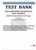
- Animal cell = Nucleus, cytoplasm, cell membrane, mitochondria and
ribosomes
- Cytoplasm is where most chemical reactions occur
- Plant cell = On top of all that a cell wall, large permanent vacuole
and chloroplasts
- Cell membrane controls what enters and leaves whereas cell wall
offers structural protection
- Bacteria have their DNA stored as a loose loop of chromosomal DNA
with plasmids (carry extra genes like resistance)
Specialised cells:
- Cells which differentiate to have specific structures for specific
functions
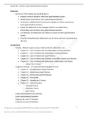
- Sperm cell: acrosome (enzymes to digest egg), flagellum,
streamlined, lots mitochondria
- Egg cell – carries female DNA, nutrients until embryo receives blood
supply
- Ciliated epithelial–line surfaces of organs and some have cilia (hairs)
to waft away dirt and pathogens
Microscope:
- Light – light rays and live specimen
- Electron – use electrons so higher resolution and magnification but
black and white and cant use live specimen
- To prep slide: water drop on clean slide, thin slice of specimen on
top, iodine drop (stain) and then cover slip
Drawing in biology: Use HB pencil, title diagram, state magnification and
title and no overlapping of lines
, Total magnification = eyepiece x objective
Magnification = image / real size
Enzymes:
- Biological catalyst which increase rate of reaction without being
used up or changed by the reaction
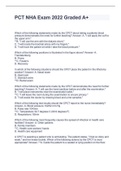
- Active site on enzyme is the bit where substrate binds to and then
the substrate is broken up into two products
- Active site shape is specific to a substrate
- Too high temp can denature active site and stop the enzyme
working but optimum temp is where the substrate has max kinetic
energy (more collisions) without denaturing active site
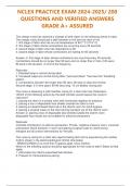
- Substrate concentration increases rate of reaction as more of the
enzyme active sites will be full until a the point where all sites are
full
- Enzymes have an optimum pH where either side of the optimum the
active site denatures
- Test how pH impacts amylase rate by dropping iodine into every well
on a spotting tile and taking a drop of an amylase with starch
solution every 30 seconds and once the amylase has broken down
all the starch the spotting tile will stay orange rather than going
black. Repeat at different pH and take times for all starch to be
broken down
- Carbohydrase’s break carbohydrates down, lipases break lipids into
fatty acids and glycerol
- Test for reducing sugars by adding benedicts reagent to food in a
test tube (in water bath) and colour spectrum goes from blue to
green to orange to red depending how much reducing sugar is
present
- Test for starch by adding iodine and if starch is present it will go
black from orange
- Test for protein by adding biuret solution to food and if protein is
present colour will change from blue to purple
Cell structure:
- Prokaryotic no nucleus
- Eukaryotic nucleus
- Animal cell = Nucleus, cytoplasm, cell membrane, mitochondria and
ribosomes
- Cytoplasm is where most chemical reactions occur
- Plant cell = On top of all that a cell wall, large permanent vacuole
and chloroplasts
- Cell membrane controls what enters and leaves whereas cell wall
offers structural protection
- Bacteria have their DNA stored as a loose loop of chromosomal DNA
with plasmids (carry extra genes like resistance)
Specialised cells:
- Cells which differentiate to have specific structures for specific
functions
- Sperm cell: acrosome (enzymes to digest egg), flagellum,
streamlined, lots mitochondria
- Egg cell – carries female DNA, nutrients until embryo receives blood
supply
- Ciliated epithelial–line surfaces of organs and some have cilia (hairs)
to waft away dirt and pathogens
Microscope:
- Light – light rays and live specimen
- Electron – use electrons so higher resolution and magnification but
black and white and cant use live specimen
- To prep slide: water drop on clean slide, thin slice of specimen on
top, iodine drop (stain) and then cover slip
Drawing in biology: Use HB pencil, title diagram, state magnification and
title and no overlapping of lines
, Total magnification = eyepiece x objective
Magnification = image / real size
Enzymes:
- Biological catalyst which increase rate of reaction without being
used up or changed by the reaction
- Active site on enzyme is the bit where substrate binds to and then
the substrate is broken up into two products
- Active site shape is specific to a substrate
- Too high temp can denature active site and stop the enzyme
working but optimum temp is where the substrate has max kinetic
energy (more collisions) without denaturing active site
- Substrate concentration increases rate of reaction as more of the
enzyme active sites will be full until a the point where all sites are
full
- Enzymes have an optimum pH where either side of the optimum the
active site denatures
- Test how pH impacts amylase rate by dropping iodine into every well
on a spotting tile and taking a drop of an amylase with starch
solution every 30 seconds and once the amylase has broken down
all the starch the spotting tile will stay orange rather than going
black. Repeat at different pH and take times for all starch to be
broken down
- Carbohydrase’s break carbohydrates down, lipases break lipids into
fatty acids and glycerol
- Test for reducing sugars by adding benedicts reagent to food in a
test tube (in water bath) and colour spectrum goes from blue to
green to orange to red depending how much reducing sugar is
present
- Test for starch by adding iodine and if starch is present it will go
black from orange
- Test for protein by adding biuret solution to food and if protein is
present colour will change from blue to purple