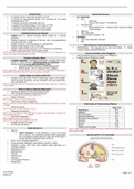
Bradley Kadou Anatomy Summary
Cell Biology:
Eukaryotes + Prokaryotes:
- Genetic Material (DNA) - Plasma Membrane (phospholipid bilayer with embedded proteins)
- Cytoplasm (semifluid matrix – doesn’t fill nucleus in eukaryotes – contains chemicals for cell functioning) -
Ribosomes (Ribonucleoprotein particles. Small in bacteria, larger in eukaryotes) –small and large subunit.
Prokaryotes:
DNA in non-membrane bound nucleoid
Cell wall is peptidoglycan (Sugars and proteins)
Smaller
Simple DNA (Double stranded ring/loop) (Small circles of DNA –Plasmids- involved in antibiotic resistance)
Eukaryotes:
DNA in membrane-bound nucleus
Cell wall is cellulose (in plant cells)
Larger
Complex DNA
Membrane bound
Prokaryotes:
Peptidoglycan can be used to divide bacteria into two groups:
1) Thick single layered wall, gram+ (retaining violet stain) 2) Multilayered cell wall, gram- (Not retaining violet
stain)
Gram staining can also be affected by lipopolysacchiride outer membrane in some bacteria WHICH affects
effectiveness of some antibiotics.
Flagella: -Hairlike projections, flagellin protein, movement, feeding.
Pili: -Shorter than flagella, attachment to substrates and to each other during conjugation (exchange of genetic
material)
Internal membranes: Invagination of the plasma membrane (cleavage)
Eukaryotes:
Outer Membrane:
Animal cells- Plasma membrane
,Plant cells- cellulose + polysaccharides and proteins
Internal structure:
Membrane bound (compartmentalization{like stories in a house, layers with differing environments}; several
processing occurring simultaneously by creating different environments) [Each membrane around an organelle is
suited to the function of that organelle]
Internal protein scaffold: extends through cytoplasm; provides support. (Like pillars of a building, internal
skeleton)
Nucleus- Double membrane bound [DNA; histone proteins and nucleolus]
Ribosomes- responsible for protein synthesis (Translation), assembled in nucleolus.
The nucleolus is the nuclear subdomain that assembles ribosomal subunits in eukaryotic cells.
Endoplasmic reticulum (ER);
-Rough (has ribosomes; protein synthesis) [proteins synthesized on surface, new protein passes into lumen,
protein vesicle buds of and passes to golgi]
-Smooth (no ribosomes; lipid synthesis) [Testosterone production and detox of liver]
Golgi Bodies- stacks of membrane, modification or proteins (Proteins and micromolecules are packaged and
sent on) (Stacks of cisternae) (Transface-outward, cisface-inward)
Lysosomes- Single membrane; breakdown of biomolecules
Vacuoles- Single membrane ; animal cells: storage; plant cell: maintain turgor pressure)
Mitochondria- Site of cell respiration (ATP synthesis), double membrane, intermembrane space and matrix. Inner
membrane folded to increase surface area (cristae)
Chloroplasts- photosynthesis. (Three membrane systems: Double enclosing membrane, inner membrane forms
thylakoids, chlorophyll in thylakoids)
Peroxisomes- breakdown of h2o2, single membrane with powerful enzymes, formed by golgi.
ENDOMEMBRANE SYSTEM (Chain): ER, Lysosomes, Vacuoles – transport to and fro from cytoplasm and
nucleus. Divides cell into compartments, channels passage of molecules in cell, provides surfaces for protein and
lipid synthesis.
Evidence for Endosymbiosis: Chloroplasts and Mitochondria have DNA. Form following function. Double
membrane bound, reproduce like bacteria.
Cell Membranes:
Plasma membrane is a lipid bilayer containing proteins, phospholipids and cholesterol. It is selectively permeable
and transports molecules using either active or passive transport.
It contains a 3-carbon glycerol (hydrophilic) attached to a fatty acid chain (hydrophobic)
, In water, the non-polar fatty acid chains attract each other to form a bilayer. This means that water-soluble
substances cannot pass through the bilayer. It must therefore contain ‘leaky’ channels to transport molecules in
and out of the cell.
Membrane proteins are amphipathic (have both hydrophilic and hydrophobic functions).
Primary structure: Amino acid chains joined by peptide bonds.
Secondary structure: Alpha-helices and Beta-pleated sheets.
Tertiary structure: 3D folding
Quaternary structure: association of 2 or more protein subunits to make 1 functional protein.
DNA Replication
5-carbon sugar molecule (Deoxyribose) (ribose in RNA)
A phosphate group
Nitrogenous Base (adenine, cytosine, guanine, *thymine) (*Uracil in RNA)
5’-3’
AT (double bond) –CG (Triple bond)
TC-pyrimidine, single ring
AG-purine, double ring
Helicase splits DNA strand into a leading and lagging strand.
DNA polymerase adds matching nucleotides. (It needs a starter, RNA primase)
Lagging strand is copied in segments (Okazaki Fragments) because it goes from 3’-5’ and DNA polymerase can
only copy from 5’-3’. These fragments then get joined by DNA Ligase.
Protein Synthesis
Begins in the nucleus, transcription unit.
RNA Polymerase copies the DNA sequence to mRNA.
mRNA moves to Ribosomes (proteins + rRna) to binding sites.
tRNA has an amino acid on one side and 3 nitrogenous bases (Codon) on the other end.
It’s matching anti-codon (UAC) attaches to the mRNA . Amino Acids join to form proteins by peptide bonds to
create a polypeptide chain.
Start codon = AUG (methionine)
3 stop codons (UAA, UAG, UGA)
Initiation- Elongation - Termination