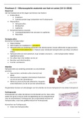
Practicum 2 – Microscopische anatomie van hart en vaten (13-11-2018)
Bouw hart
Het hart bestaat ook uit drie lagen (van binnen naar buiten):
• Endocardium:
• Endotheel.
• Subendocardiale laag: bindweefsel met Purkinjevezels.
• Myocardium:
• Hartspiercellen.
• Bindweefsel.
• Capillairen.
• Epicardium/Lamina visceralis:
• Lozmazig bindweefsel met zenuwen en capillairen.
• Mesotheel.
Hartspiercellen
Kenmerken hartspiercellen:
• Dwarsstreping.
• Geen fusatie van myoblasten (= syncytium).
• Hartspiercellen zijn onderling verbonden met desmonsomen, fasciae adherentes en gap junctions.
→ Glanslijnen/Intercalated discs (= Plaatsen waar individuele hartspiercellen aan elkaar verankerd
zitten).
• Hartspiercellen zijn niet cylindrisch, maar vertakte structuren. → Alle hartspiercellen trekken
tegelijkertijd samen.
• De celkern ligt in het midden van de cel.
• Bevatten veel mitochondriën.
• Functie: contraheren.
Glanslijn
Hartspiercellen zijn onderling verbonden via verschillende
junctions:
• Gap junctions: verbinden cellen chemisch met
elkaar.
• Desmonsomen: koppelen intermediaire filamenten
van de cel aan het celmembraan.
• Adherens junctions/Fasciale adherentes: koppelen
actine filamenten van de cel aan het celmembraan.
De glanslijnen bestaan uit uitstulpingen van het uiteinde van de ene hartspiercel in de andere hartspiercel.
Purkinjevezels
Purkinjevezels zijn cellen die behoren tot de Bundel van His. Deze cellen
spelen een grote rol bij de snelle voortgeleiding van het actiepotentiaal
over de ventrikelwand. Purkinjevezels zijn oorspronkelijk ontstaan uit
hartspiercellen, maar later gedifferentieerd tot Purkinjevezels.
Kenmerken van Purkinjevezels:
• Groter dan hartspiercellen.
1