Case 1 introduction to clinical child neuropsychology
1. Define the different stages of brain development (prenatal, postnatal, adolescence);
what changes can be distinguished in each period?
- Between birth and adulthood the human brain quadruples in size, increasing from
around 400 g at birth to 1500 g at maturity in early adulthood, peaking between 18 and
30 years and then commencing a gradual decline. The postnatal increase in brain
weight is largely due to differentiation, growth, and maturation of existing neurons,
including elaboration of dendrites and synapses, and
ongoing myelination.
Prenatal development
- Prenatal development is primarily concerned with the
structural formation of the CNS, and is thought to be largely
genetically determined.
- Grow spurts: The earliest of these spurts has been
documented around 24-25 weeks gestation, coinciding with
the completion of neuronal generation. A further spurt
occurs during the first year of life due to dendritic and
synaptic development and myelination. Later spurts have
been identified between 7 and 9 years of age, and a final
spurt around 16-19 years. A disruption to development
during a growth spurt may be particularly detrimental to
ongoing development, causing a cessation of development or altering the
course of development.
- there is a hierarchical progression within. the CNS, with cerebellar/brain
stem areas maturing first, followed by posterior areas, and lastly anterior
regions, particularly the frontal cortex. This hierarchical development is
argued to progress in spurts, representing the ongoing elaboration of the
system
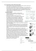
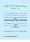
- The fertilised cell experiences rapid cell division, resulting in the
formation of a cluster of cells which quickly becomes the embryonic disc.
The embryonic disc comprises three layers which later form specific
organic systems within the human body. The nervous system emerges-via
a process of neurolation, from the outer layer, or ectoderm, of the
embryonic disc, which folds in on itself and forms a tube. The initial
stages of neurolation are thought to commence during the second week of
gestation. In the third week of gestation, the neural plate becomes visible
as a thickened area of the ectoderm. Gradually a longitudinal neural
groove begins to form, and is flanked by two edges or neural folds. These
neural folds deepen and fold until they fuse,
creating a hollow cylindrical structure, the
neural tube, by week 4 of gestation
1
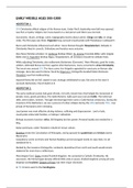
, - - In contrast to the active role of the neuron, glial cells
play a more supportive and nutrient role within the nervous
system, supporting the neurons,. enabling regeneration of
damaged neurons, producing scar tissue to occupy damage sites
and transporting nutrients from nerve cells. There are nine
times as many glial cells within the nervous system as there are
neurons. Glial cells are relatively immature in the early stages
of development and continue to generate with the increased
maturity of the CNS.
- Cell proliferation: Each of the three layers of embryonic disc is
destined to form a major organic system. The inner layer
(endoderm) will form the internal organs, including the
digestive and respiratory systems, whereas the skeletal and
muscular structures will develop from the middle layer, or
mesoderm. The outer layer, or ectoderm, eventually forms the
nervous system and the skin surface. Neurogenesis occurs within the neural tube. It
begins early in gestation, around day 40, and is virtually complete by 6 months
gestation, with the exception of a small number of cerebellar and hippocampal cells
that continue to divide even after birth. Cell proliferation occurs within the neural tube
in the germinal matrix, in the ventricular proliferative zone, and cells then migrate
from there to predetermined locations within the nervous system. The process appears
to be precisely regulated so that appropriate numbers of cells are formed at
predetermined times and in well-defined regions
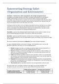
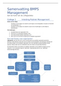
- Cell migration: At the completion of the cell proliferation stage, but not before 6
weeks gestation, the neuroblasts formed within the neural tube begin to move, or
migrate, to their permanent locations. Two forms of migration have been identified.
Passive migration, or cell displacement, occurs when cells are simply pushed away
from where they originated by other cells that were generated more recently. In
consequence these older cells are moved gradually towards the surface of the brain,
whereas newer cells take up more internal positions. The second form, active
migration, occurs when younger cells "overtake" the migrational activity of older cells
them to external regions including the cerebral cortex. Cells migrate along these glial
fibres to genetically predetermined regions of the brain.
- Cell differentiation: Once neurons have migrated they begin the process of
differentiation, which occurs as four simultaneous processes: (1) development of cell
bodies; (2) selective cell death; (3) dendritic and axonal growth and (4) formation of
synaptic connections. Simultaneously glial cells differentiating, with oligodendrocytes
2
1. Define the different stages of brain development (prenatal, postnatal, adolescence);
what changes can be distinguished in each period?
- Between birth and adulthood the human brain quadruples in size, increasing from
around 400 g at birth to 1500 g at maturity in early adulthood, peaking between 18 and
30 years and then commencing a gradual decline. The postnatal increase in brain
weight is largely due to differentiation, growth, and maturation of existing neurons,
including elaboration of dendrites and synapses, and
ongoing myelination.
Prenatal development
- Prenatal development is primarily concerned with the
structural formation of the CNS, and is thought to be largely
genetically determined.
- Grow spurts: The earliest of these spurts has been
documented around 24-25 weeks gestation, coinciding with
the completion of neuronal generation. A further spurt
occurs during the first year of life due to dendritic and
synaptic development and myelination. Later spurts have
been identified between 7 and 9 years of age, and a final
spurt around 16-19 years. A disruption to development
during a growth spurt may be particularly detrimental to
ongoing development, causing a cessation of development or altering the
course of development.
- there is a hierarchical progression within. the CNS, with cerebellar/brain
stem areas maturing first, followed by posterior areas, and lastly anterior
regions, particularly the frontal cortex. This hierarchical development is
argued to progress in spurts, representing the ongoing elaboration of the
system
- The fertilised cell experiences rapid cell division, resulting in the
formation of a cluster of cells which quickly becomes the embryonic disc.
The embryonic disc comprises three layers which later form specific
organic systems within the human body. The nervous system emerges-via
a process of neurolation, from the outer layer, or ectoderm, of the
embryonic disc, which folds in on itself and forms a tube. The initial
stages of neurolation are thought to commence during the second week of
gestation. In the third week of gestation, the neural plate becomes visible
as a thickened area of the ectoderm. Gradually a longitudinal neural
groove begins to form, and is flanked by two edges or neural folds. These
neural folds deepen and fold until they fuse,
creating a hollow cylindrical structure, the
neural tube, by week 4 of gestation
1
, - - In contrast to the active role of the neuron, glial cells
play a more supportive and nutrient role within the nervous
system, supporting the neurons,. enabling regeneration of
damaged neurons, producing scar tissue to occupy damage sites
and transporting nutrients from nerve cells. There are nine
times as many glial cells within the nervous system as there are
neurons. Glial cells are relatively immature in the early stages
of development and continue to generate with the increased
maturity of the CNS.
- Cell proliferation: Each of the three layers of embryonic disc is
destined to form a major organic system. The inner layer
(endoderm) will form the internal organs, including the
digestive and respiratory systems, whereas the skeletal and
muscular structures will develop from the middle layer, or
mesoderm. The outer layer, or ectoderm, eventually forms the
nervous system and the skin surface. Neurogenesis occurs within the neural tube. It
begins early in gestation, around day 40, and is virtually complete by 6 months
gestation, with the exception of a small number of cerebellar and hippocampal cells
that continue to divide even after birth. Cell proliferation occurs within the neural tube
in the germinal matrix, in the ventricular proliferative zone, and cells then migrate
from there to predetermined locations within the nervous system. The process appears
to be precisely regulated so that appropriate numbers of cells are formed at
predetermined times and in well-defined regions
- Cell migration: At the completion of the cell proliferation stage, but not before 6
weeks gestation, the neuroblasts formed within the neural tube begin to move, or
migrate, to their permanent locations. Two forms of migration have been identified.
Passive migration, or cell displacement, occurs when cells are simply pushed away
from where they originated by other cells that were generated more recently. In
consequence these older cells are moved gradually towards the surface of the brain,
whereas newer cells take up more internal positions. The second form, active
migration, occurs when younger cells "overtake" the migrational activity of older cells
them to external regions including the cerebral cortex. Cells migrate along these glial
fibres to genetically predetermined regions of the brain.
- Cell differentiation: Once neurons have migrated they begin the process of
differentiation, which occurs as four simultaneous processes: (1) development of cell
bodies; (2) selective cell death; (3) dendritic and axonal growth and (4) formation of
synaptic connections. Simultaneously glial cells differentiating, with oligodendrocytes
2