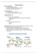
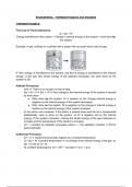
Patterns of Inheritance
Asexual reproduction
● A single individual is the parent
● All genetic information is passed to the offspring
● Produces exact genetic copies “clones”
Sexual reproduction
● Two parents produce an offspring
● Combination of genes (50%) from each parent
Haploid vs. Diploid cells
● Diploid
○ Cell with two chromosome sets
○ Sexual reproducing species contain both haploid and diploid cells
○ Non-sex cells
○ 2n=46
● Haploid
○ Cells with single chromosome sets
○ Sex cells
○ n=23
Sexual life-cycle: Fertilization and meiosis must alternate to help maintain a constant
number of chromosomes in each species to the next
● Meiosis maintains a normal diploid number of chromosomes (46)
● Fertilization maintains a normal haploid number of chromosomes (26)
● When two haploid cells come together a zygote is created which then divides
using mitosis
, Stages of meiosis: Two consecutive cell divisions
1. Prophase I Longest stage of meiosis. Chromatin shortens and thickens
becoming chromosomes. Centrioles move towards the poles. Rather than
scattering like in mitosis.
● Synaptonemal complex forms between the homologous chromosomes holding
them in synapsis.
● When the members of the chromosomes pair up to find each other, the pairing
of homologous chromosomes is called synapsis.
● Once chromosomes synapsed, the arms may cross over at varying points. The
crossover causes genes to be shuffled.
● The nucleus and nuclear membrane disintegrate. Spindles and aster rays form,
microtubules attach to kinetochores, centrosome and homogolous movement
● Chiasmata: A structure that holds homologous pairs together as spindles form
2. Metaphase I Paired chromosomes line up at the metaphase plate, each
chromatid is attached to kinetochore.
3. Anaphase I Each part of homologous chromosomes separate from their
partner due to protein breakdown, each goes to opposite poles. Meanwhile,
cohesion persists, chromatids sticking together
4. Telophase I/ Cytokinesis The cytoplasm divides with complete haploid set.
5. Prophase II New spindle fibres and astral rays form. Centrioles double and
move to poles.
6. Metaphase II Chromatids line up at the equator in a single file. Kinetochores
attach to spindles.
7. Anaphase II Chromatids separate and move to opposite poles due to the
breakdown of proteins holding the chromatids together
8. Telophase II/ Cytokinesis Cytoplasm divides. Spindle astral rays disappear.
nuclear membrane and nucleolus reform, chromosomes decondense.
4 daughter cells created with different genetic material