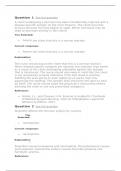
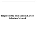
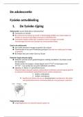
Vit D and Ca Absorption PTH
Low Ca
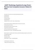
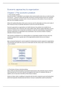
Pre-renal* Renal Post-renal Low PO4
Caused by any condition leading to decreased renal perfusion. Caused by direct kidney damage. Caused by any condition that results in bilateral obstruction of UV-B
Absorbed from diet
urinary flow from renal pelvis to bladder. Calcitriol
Cholecalciferol (Vit D3) Calcifediol
~ 60% cases ~ 35% cases (inactive) (active)
~ 5% cases
1. Hypovolemia 1. Vascular (TTP, malignant htn, vasculitis, thrombotic
A. Absolute (volume depletion) microagniopathy, cholesterol emboli, large vessel dz) Small intestine Liver Kidney
1. Congenital malformations (posterior urethral valves)
• Hemorrhage, GI loss, skin loss, renal loss 2. Glomerulonephritis (RPGN) 2. Acquired obstructions
B. Effective (decreased circulating volume) • Cholesterol emboli Small intestine Enterocyte Blood vessel
• BPH
• CHR, cirrhosis, 3rd spacing, sepsis, shock • HUS • Iatrogenic/catheter-associated (duodenum, proximal jejunum)
2. Vascular (large vessel): renal a stenosis, vasculitis, • DIC • Tumour/LNs
dissection, VT • Malignant htn • Stones (nephrolithiasis) Mechanisms of Ca absorption:
3. Vascular (renal vasoconstriction): NSAIDs, ACEI, 3. Acute interstitial nephritis (AIN) • Clotting (vascular) 1. Active transcellular process restricted to l
rio
cyclosporin/tacrolimus, contrast • Drug-induced (penicillin, b-lactam, PPI, quinolones) 3. Neurogenic bladder (MS, spinal cord lesions, etc.) duodenum and proximal jejunum lcit
Ca VD
• Infectious (pyelo, legionella, TB) 4. Drugs: Anticholinergics = retention 2. Passive process, length of intestine R
TF
INVESTIGATIONS • Infiltrative (sarcoid, lymphoma, leukemia)
• ↓ BP, ↑ HR, orthostatic changes • Autoimmune (Sjogren’s, SLE) PRESENTATION/INVESTIGATIONS
• ↑ [urea] >> ↑ [Cr] 4. Acute tubular necrosis* (ATN) Ca transporter
• Known solitary kidney
• Urine [Na] < 20 mmol/L • Ischemia (progressive from prerenal; sepsis MC) • Older man
• Urine osmolality > 500 mOsm/kg • Toxins (drugs, Hgb/myoglobin) • Recent retroperitoneal surgery
• FeNa < 1% • Contrast • Anuria Ca2+
• Palpable bladder
MANAGEMENT INVESTIGATIONS • US with hydronephrosis Ca transporter
Calbindin D
• Fluids to optimize volume status and cardiac performance • Systemic features: anemia, thrombocytopenia, htn, volume
(NS, albumin, blood/plasma) Ca2+
overload MANAGEMENT
• Hold meds if possible • Casts: • Tx obstruction cause: structural (stone, strictures) vs
• GN – RBC function (neuropathy) Ca2+
ATP
• AIN – WBC • Foley cath, indwelling bladder cath, nephrostomy, Ca pump Ca2+
• ATN – pigmented granular (muddy brown) stenting
MANAGEMENT
VDR = vitamin D receptor/calcitriol receptor (TF)
• Tx infection
• Optimize lytes
• D/c nephrotoxic drugs
• Fluids to optimize volume status
• Supportive are
• +/- steroids/immunosuppressants
Nephrology Summary Notes
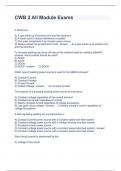
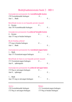
Minimal change Membranous GN FSGS Membranoproliferati Nodular GS Fluid Compartments ECF 1/3
ve GN
Proposed T cell-mediated Immune complexes Injury to podocytes Immune complexes Marked mesangial
pathophysiology cytokine injury of in blood-gas barrier in blood-gas barrier expansion H2O H2O
podocytes
Interstitial
2/3
Secondary causes Hodgkin’s lymphoma HBV, SLE, solid Reflux nephropathy, HCV, malaria, SLE, DM, amyloidosis ICF Na
tumours HIV, HBV, obesity, leukemia, lymphoma, 2/3 H2O
sickle cell disease shunt nephritis
Capillary
H2O H2O
Drug causes NSAIDs, Lithium Gold, penicillamine Heroin Intravascular
1/3 Na
Therapy Steroids Reduce bp, ACEi, Steroids, ACEi/ARB ASA, ACEi, Tx underlying cause H2O
steroids for proteinuria dipyridamole
(controversial)
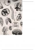
, Renal Tubule
Glomerulus
Proximal convoluted tubule Distal convoluted tubule
Late
Renal Corpuscle Early
Renal Cortex
PTH Thiazide diuretics
Bowman’s Capsule Na
Glucose Na
Na
HCO3 Cl H+ Cl
Urea Ca K ADH
Osmotic diuretics
H2O
325 mOsm/L Aldosterone
NH4
Medications Na
Cl
300 mOsm/L K
Ascending limb
Loop diuretics Na, H2O
Renal Medulla
H+, K
H2O
600 mOsm/L K-sparing diuretics
Na
Descending limb Cl
Collecting duct
1200 mOsm/L
Loop of Henle Urine to ureter
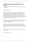
,Diuretics Summary
Types MOA Indications Dosing SE Contraindications
Thiazides Hydrochlorothiazide, ↓ Na reabsorption in DCT Htn (1st line essential htn), HCTZ: Hypotension, Sulfa allergy,
chlorthiazide edema, idiopathic Htn: 12.5-25 mg PO od hypokalemia, polyuria pregnancy
hypercalciuria and stones (max 50 mg/d)
Edema: 25-100 mg PO od
Stones: 25-100 mg PO od
Loop diuretics Furosemide (Lasix) Blocks luminal Na/K/Cl CHF, Edema: 20-80 mg IV/IM/PO Hypovolemia, Hypovolemia,
transporter in thick pulmonary/peripheral q6-8h (max 600 mg/d) hypokalemia, metabolic hypokalemia
ascending limb edema, Htn Htn: 20-80 mg/d PO od/BID alkalosis
↓ Na, K, Cl reabsorption
K-sparing/ Spironolactone, Antagonizes aldosterone R Htn, CHF, hypokalemia Spironolactone: Edema, hyperkalemia, Renal insufficiency,
Aldosterone eplenerone to ↓ Na at collecting duct Htn: 25-200 mg/d od/BID gynecomastia hyperkalemia,
antagonists pregnancy
Osmotic Mannitol (Osmitol) Non-reabsorbable solute Decrease intracranial and Mannitol: Transient volume Anuria, active cranial
Glycerol increases osmotic pressure intraocular pressure ↓ICP: 0.25-2 g/kg IV over expansion bleeding
Urea of filtrate; inhibits H2O RF/edematous states 30-60 mins Electrolyte
reabsorption at PCT and abnormalities
collecting duct
, Measurement of Renal Function
Renal blood panel
Note: Increased muscle mass = increased creatine
• GFR is estimated: Test Association
• Clinically using serum [Cr]
• Metabolite of creatine (intermediate in muscle metabolism)
• Cr is freely filtered at glomerulus with little tubular reabsorption Lytes: Na, K, Cl, HCO3 Renal dz can contribute to electrolyte disturbance
• At steady state [Cr] = 1/CrCl therefore sudden insult (AKI) will not be reflected in Cr
immediately
• GFR/day = (urine [Cr] x 24h urine volume )/(plasma [Cr]) PO4 ↑ plasma [ ] in renal dz
• Increased GFR in pregnancy
• Gold standard to estimate is using inulin clearance
Ca ↓ plasma [ ] in renal dz
• Cystatin C = protease completely filtered by glomerulus and not affected by muscle mass
• Urea = end product of protein metabolism Albumin ↓ plasma [ ] in renal dz (nephrotic syndrome)
• Plasma [urea] reflect renal function
• High urea independent of renal function: high protein diet, volume depletion (prerenal
azotemia), GI hemorrhage, sepsis, catabolic state, corticosteroid/cytotoxic agents Urea ↑ plasma [ ] suggests renal dz or decreased blood flow
• Low urea independent of renal function: low protein diet, liver dz to kidneys or urine obstruction
Creatinine (Cr) ↑ plasma [ ] suggests renal dz or decreased blood flow
to kidneys or urine obstruction
HbA1c ↑ in DM; common cause of renal dz
Cystatin C ↑ plasma [ ] suggests renal dz (low GFR)
eGFR Calculated from blood Cr
eGFR < 60 mL/min = renal damage
eGFR < 15 mL/min = renal failure
Anion gap Help determine cause of metabolic acidosis
Low Ca
Pre-renal* Renal Post-renal Low PO4
Caused by any condition leading to decreased renal perfusion. Caused by direct kidney damage. Caused by any condition that results in bilateral obstruction of UV-B
Absorbed from diet
urinary flow from renal pelvis to bladder. Calcitriol
Cholecalciferol (Vit D3) Calcifediol
~ 60% cases ~ 35% cases (inactive) (active)
~ 5% cases
1. Hypovolemia 1. Vascular (TTP, malignant htn, vasculitis, thrombotic
A. Absolute (volume depletion) microagniopathy, cholesterol emboli, large vessel dz) Small intestine Liver Kidney
1. Congenital malformations (posterior urethral valves)
• Hemorrhage, GI loss, skin loss, renal loss 2. Glomerulonephritis (RPGN) 2. Acquired obstructions
B. Effective (decreased circulating volume) • Cholesterol emboli Small intestine Enterocyte Blood vessel
• BPH
• CHR, cirrhosis, 3rd spacing, sepsis, shock • HUS • Iatrogenic/catheter-associated (duodenum, proximal jejunum)
2. Vascular (large vessel): renal a stenosis, vasculitis, • DIC • Tumour/LNs
dissection, VT • Malignant htn • Stones (nephrolithiasis) Mechanisms of Ca absorption:
3. Vascular (renal vasoconstriction): NSAIDs, ACEI, 3. Acute interstitial nephritis (AIN) • Clotting (vascular) 1. Active transcellular process restricted to l
rio
cyclosporin/tacrolimus, contrast • Drug-induced (penicillin, b-lactam, PPI, quinolones) 3. Neurogenic bladder (MS, spinal cord lesions, etc.) duodenum and proximal jejunum lcit
Ca VD
• Infectious (pyelo, legionella, TB) 4. Drugs: Anticholinergics = retention 2. Passive process, length of intestine R
TF
INVESTIGATIONS • Infiltrative (sarcoid, lymphoma, leukemia)
• ↓ BP, ↑ HR, orthostatic changes • Autoimmune (Sjogren’s, SLE) PRESENTATION/INVESTIGATIONS
• ↑ [urea] >> ↑ [Cr] 4. Acute tubular necrosis* (ATN) Ca transporter
• Known solitary kidney
• Urine [Na] < 20 mmol/L • Ischemia (progressive from prerenal; sepsis MC) • Older man
• Urine osmolality > 500 mOsm/kg • Toxins (drugs, Hgb/myoglobin) • Recent retroperitoneal surgery
• FeNa < 1% • Contrast • Anuria Ca2+
• Palpable bladder
MANAGEMENT INVESTIGATIONS • US with hydronephrosis Ca transporter
Calbindin D
• Fluids to optimize volume status and cardiac performance • Systemic features: anemia, thrombocytopenia, htn, volume
(NS, albumin, blood/plasma) Ca2+
overload MANAGEMENT
• Hold meds if possible • Casts: • Tx obstruction cause: structural (stone, strictures) vs
• GN – RBC function (neuropathy) Ca2+
ATP
• AIN – WBC • Foley cath, indwelling bladder cath, nephrostomy, Ca pump Ca2+
• ATN – pigmented granular (muddy brown) stenting
MANAGEMENT
VDR = vitamin D receptor/calcitriol receptor (TF)
• Tx infection
• Optimize lytes
• D/c nephrotoxic drugs
• Fluids to optimize volume status
• Supportive are
• +/- steroids/immunosuppressants
Nephrology Summary Notes
Minimal change Membranous GN FSGS Membranoproliferati Nodular GS Fluid Compartments ECF 1/3
ve GN
Proposed T cell-mediated Immune complexes Injury to podocytes Immune complexes Marked mesangial
pathophysiology cytokine injury of in blood-gas barrier in blood-gas barrier expansion H2O H2O
podocytes
Interstitial
2/3
Secondary causes Hodgkin’s lymphoma HBV, SLE, solid Reflux nephropathy, HCV, malaria, SLE, DM, amyloidosis ICF Na
tumours HIV, HBV, obesity, leukemia, lymphoma, 2/3 H2O
sickle cell disease shunt nephritis
Capillary
H2O H2O
Drug causes NSAIDs, Lithium Gold, penicillamine Heroin Intravascular
1/3 Na
Therapy Steroids Reduce bp, ACEi, Steroids, ACEi/ARB ASA, ACEi, Tx underlying cause H2O
steroids for proteinuria dipyridamole
(controversial)
, Renal Tubule
Glomerulus
Proximal convoluted tubule Distal convoluted tubule
Late
Renal Corpuscle Early
Renal Cortex
PTH Thiazide diuretics
Bowman’s Capsule Na
Glucose Na
Na
HCO3 Cl H+ Cl
Urea Ca K ADH
Osmotic diuretics
H2O
325 mOsm/L Aldosterone
NH4
Medications Na
Cl
300 mOsm/L K
Ascending limb
Loop diuretics Na, H2O
Renal Medulla
H+, K
H2O
600 mOsm/L K-sparing diuretics
Na
Descending limb Cl
Collecting duct
1200 mOsm/L
Loop of Henle Urine to ureter
,Diuretics Summary
Types MOA Indications Dosing SE Contraindications
Thiazides Hydrochlorothiazide, ↓ Na reabsorption in DCT Htn (1st line essential htn), HCTZ: Hypotension, Sulfa allergy,
chlorthiazide edema, idiopathic Htn: 12.5-25 mg PO od hypokalemia, polyuria pregnancy
hypercalciuria and stones (max 50 mg/d)
Edema: 25-100 mg PO od
Stones: 25-100 mg PO od
Loop diuretics Furosemide (Lasix) Blocks luminal Na/K/Cl CHF, Edema: 20-80 mg IV/IM/PO Hypovolemia, Hypovolemia,
transporter in thick pulmonary/peripheral q6-8h (max 600 mg/d) hypokalemia, metabolic hypokalemia
ascending limb edema, Htn Htn: 20-80 mg/d PO od/BID alkalosis
↓ Na, K, Cl reabsorption
K-sparing/ Spironolactone, Antagonizes aldosterone R Htn, CHF, hypokalemia Spironolactone: Edema, hyperkalemia, Renal insufficiency,
Aldosterone eplenerone to ↓ Na at collecting duct Htn: 25-200 mg/d od/BID gynecomastia hyperkalemia,
antagonists pregnancy
Osmotic Mannitol (Osmitol) Non-reabsorbable solute Decrease intracranial and Mannitol: Transient volume Anuria, active cranial
Glycerol increases osmotic pressure intraocular pressure ↓ICP: 0.25-2 g/kg IV over expansion bleeding
Urea of filtrate; inhibits H2O RF/edematous states 30-60 mins Electrolyte
reabsorption at PCT and abnormalities
collecting duct
, Measurement of Renal Function
Renal blood panel
Note: Increased muscle mass = increased creatine
• GFR is estimated: Test Association
• Clinically using serum [Cr]
• Metabolite of creatine (intermediate in muscle metabolism)
• Cr is freely filtered at glomerulus with little tubular reabsorption Lytes: Na, K, Cl, HCO3 Renal dz can contribute to electrolyte disturbance
• At steady state [Cr] = 1/CrCl therefore sudden insult (AKI) will not be reflected in Cr
immediately
• GFR/day = (urine [Cr] x 24h urine volume )/(plasma [Cr]) PO4 ↑ plasma [ ] in renal dz
• Increased GFR in pregnancy
• Gold standard to estimate is using inulin clearance
Ca ↓ plasma [ ] in renal dz
• Cystatin C = protease completely filtered by glomerulus and not affected by muscle mass
• Urea = end product of protein metabolism Albumin ↓ plasma [ ] in renal dz (nephrotic syndrome)
• Plasma [urea] reflect renal function
• High urea independent of renal function: high protein diet, volume depletion (prerenal
azotemia), GI hemorrhage, sepsis, catabolic state, corticosteroid/cytotoxic agents Urea ↑ plasma [ ] suggests renal dz or decreased blood flow
• Low urea independent of renal function: low protein diet, liver dz to kidneys or urine obstruction
Creatinine (Cr) ↑ plasma [ ] suggests renal dz or decreased blood flow
to kidneys or urine obstruction
HbA1c ↑ in DM; common cause of renal dz
Cystatin C ↑ plasma [ ] suggests renal dz (low GFR)
eGFR Calculated from blood Cr
eGFR < 60 mL/min = renal damage
eGFR < 15 mL/min = renal failure
Anion gap Help determine cause of metabolic acidosis