Radiologie Hals
Radio-anatomie 1e BA GNK
Beeldvormingstechnieken:
- RX: niet meer gebruikt, zeker niet voor de weke delen
- echografie: geen ioniserende stralen, eenvoudig en snel beschikbaar, goed voor opp. structuren.
onder echografie kunnen puncties uitgevoerd worden van:
- klieren - parotis - spieren - schildklier
- CT: ruim beschikbaar, goed de diepe structuren in beeld brengen, 3D, gebruikt wel ioniserende
straling.
met of zonder intraveneuze contrast toediening
met: goed onderscheid tussen vasculaire structuren, spierbundels, speekselklieren,…
zonder: beperkte indicaties
een standaard MR wordt dus zeker met contrast gedaan.
- MRI: beste manier voor weke delen, geen ioniserende stralen, beperkt beschikbaar
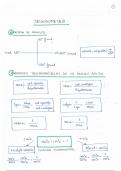
- De hals wordt opgedeeld in ‘ruimtes’ of ‘loges’ belijnd door fascia. Het compartiment is van belang
voor het bepalen van de aandoening. Het bepaalt bv. het stadium van een bepaalde tumor, de
uitzaaiing.
Opgedeeld in suprahyoid en infrahyoid.
- te werk gaan: 1) afwijking detecteren 2) kijken in welke ruimte deze ligt
3) de normale anatomische structuren van deze ruimte bekijken
4) radiologische kenmerken en klinische informatie 5) diagnose
Aero digestieve Tractus:
Farynx: - nasopharynx
(clivus – weke gehemelte)
- orofarynx
(weke gehemelte tot valleculae)
Larynx: van epiglottis tot cricoid
- overlap tussen de larynx en de hypofarynx
- nasofarynx: -
Orofarynx:
- hypofarynx:
1
, Radiologie Hals
Radio-anatomie 1e BA GNK
- Ring van Waldeyer
- Larynx:
supraglottis: van epiglottis tot ventriculus laryngi
glottis: ware stembanden
subglottis: tot cricoid kraakbeen, gaat over in trachea
- skelet: Hyoid = tongbeen
thyroid
arythenoid
cricoid
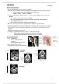
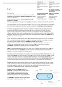
Re schildklier
Supra- en infrahyoïdale hals:
- oppervlakkige halsfascia:
direct onder de subcutis gelegen, omgeeft de platysma en de
mimische spieren. Loopt van epicranium tot aan de thorax en de
oksel.
- diepe halsfascia:
omgeven de verschillende ruimten in de hals. Geven steun
aan de spieren, organen, bloedvaten en zenuwen.
ze worden opgedeeld in 3 lagen:
- oppervlakkige - middelste - diepe
Belangrijkste compartimenten vd hals:
2
Radio-anatomie 1e BA GNK
Beeldvormingstechnieken:
- RX: niet meer gebruikt, zeker niet voor de weke delen
- echografie: geen ioniserende stralen, eenvoudig en snel beschikbaar, goed voor opp. structuren.
onder echografie kunnen puncties uitgevoerd worden van:
- klieren - parotis - spieren - schildklier
- CT: ruim beschikbaar, goed de diepe structuren in beeld brengen, 3D, gebruikt wel ioniserende
straling.
met of zonder intraveneuze contrast toediening
met: goed onderscheid tussen vasculaire structuren, spierbundels, speekselklieren,…
zonder: beperkte indicaties
een standaard MR wordt dus zeker met contrast gedaan.
- MRI: beste manier voor weke delen, geen ioniserende stralen, beperkt beschikbaar
- De hals wordt opgedeeld in ‘ruimtes’ of ‘loges’ belijnd door fascia. Het compartiment is van belang
voor het bepalen van de aandoening. Het bepaalt bv. het stadium van een bepaalde tumor, de
uitzaaiing.
Opgedeeld in suprahyoid en infrahyoid.
- te werk gaan: 1) afwijking detecteren 2) kijken in welke ruimte deze ligt
3) de normale anatomische structuren van deze ruimte bekijken
4) radiologische kenmerken en klinische informatie 5) diagnose
Aero digestieve Tractus:
Farynx: - nasopharynx
(clivus – weke gehemelte)
- orofarynx
(weke gehemelte tot valleculae)
Larynx: van epiglottis tot cricoid
- overlap tussen de larynx en de hypofarynx
- nasofarynx: -
Orofarynx:
- hypofarynx:
1
, Radiologie Hals
Radio-anatomie 1e BA GNK
- Ring van Waldeyer
- Larynx:
supraglottis: van epiglottis tot ventriculus laryngi
glottis: ware stembanden
subglottis: tot cricoid kraakbeen, gaat over in trachea
- skelet: Hyoid = tongbeen
thyroid
arythenoid
cricoid
Re schildklier
Supra- en infrahyoïdale hals:
- oppervlakkige halsfascia:
direct onder de subcutis gelegen, omgeeft de platysma en de
mimische spieren. Loopt van epicranium tot aan de thorax en de
oksel.
- diepe halsfascia:
omgeven de verschillende ruimten in de hals. Geven steun
aan de spieren, organen, bloedvaten en zenuwen.
ze worden opgedeeld in 3 lagen:
- oppervlakkige - middelste - diepe
Belangrijkste compartimenten vd hals:
2