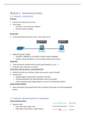
2.4: Perception
Problem 1: The Eye
The Eye
- roughly spherical with a diameter of about 24 mm
Þ optic axis – imaginary diameter from the front to the back
of the eye, passing through the centre of the lens
o each eye points in the direction defined by the optic
axis
Þ Membranes
o sclera – outer membrane à a tough protective
covering whose visible portion is the white of the eye
and the transparent cornea at the front of the eye
o choroid – middle membrane à lines the interior of the
sclera and contains most of the blood vessels that
supply the inside of the eye with oxygen and nutrients
o retina – inner membrane à made up of neurons,
including the receptors that convert the light entering
the eye into neural signals
Þ cornea – transparent membrane at the front of the eye
o light enters the eye by first passing through the cornea
which sharply refracts (bends) the light
o performs most of the focusing light on the retina process
o rigid; cannot adjust how much light passing through is refracted
Þ pupil – an opening in the middle of the iris through which light enters the eye
o diameter: 2-8mm
o when pupil gets smaller à it constricts
o when pupil gets larger à it dilates
Þ iris – coloured part of the eye with an opening in the middle (the pupil)
o pupillary reflex – controls the size of the pupil by contracting and relaxing , mainly in
response to the intensity of light entering the eye
• intense light à iris contracts àsmaller pupil à reduces the amount of light that
can enter the eye
• dim light à iris relaxes à larger pupil à increases the amount of light that can
enter the eye
Þ Chambers
o anterior chamber – the space between the cornea and the iris
• filled with aqueous humor – a clear thin fluid
o posterior chamber – the space between the iris and the lens
• filled with aqueous humor – a clear thin fluid
o vitreous chamber – the main interior portion of the eye
• filled with vitreous humor – a clear, gel-like fluid
è the aqueous & vitreous humor slightly refract light, O2 and nutrients
Þ lens – a transparent structure that further refracts the light, to ensure that light focuses
properly on the retina
o focal length – the distance from the lens at which the image of an object is in focus
when the object is far away from the lens
, • strong lens à refracts light sharply à it is relatively thick, rounded, has a short
focal length
o zonule fibres – fibres that connect the lens to the choroid; they pull on the lens to
change its shape
o ciliary muscles – tiny muscles attached to the choroid; they relax and contract to
control how the choroid pulls on the zonule fibres to change the shape of the lens à
increase the focusing power of the lens
Þ optic disk (blind spot) – location on the retina where the axons of RGCs exit the eye
through the optic disk
o axons of the RGC come together at the optic disk and exit the eye in a bundle (the optic
nerve)
Þ optic nerve – formed by the bundling together of axons of RGCs; it exits the eye through
the optic disk
Þ fovea – a region at the centre of the retina where the light from objects at the centre of our
gaze strikes the retina
o contains no rods
o very high density of cones
Þ accommodation – the adjustment of the shape of the lens to focus on objects at different
distances from the eye
o ciliary muscles: relaxed à the choroid can pull on the zonule fibres
• this stretches the lens = relatively thin, flat shape = relatively weak lens with a
relatively long focal length, appropriate for focusing light from distant objects
o ciliary muscles: contract à they oppose the pull by the choroid on the zonule fibres
• lens isn't stretched as much = thicker, more rounded shape = a stronger lens with
a shorter focal length, appropriate for focusing light from nearer objects
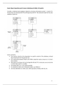
Anatomy of the Retina
Þ made up of several different classes of neurons – each class performs a distinct function
Þ photoreceptors – retinal neurons (rods and cones) that transduce light into neural signals;
send signals to bipolar cells; send to and receive signals from horizontal cells
o rods – provide black-and-white vision in dim light
o cones – provide high-acuity colour vision in bright light
Þ pigment epithelium – a layer of cells attached to the choroid; photoreceptors are embedded
in it
o receive nourishment from it
Þ retina à structured in layers à nuclear layers – the three main layers (outer nuclear, inner
nuclear, ganglion cell layers); because they contain the nuclei of various types of retinal
neurons
1. outer nuclear layer – consists of the photoreceptors (not including their inner and
outer segments)
2. inner nuclear layer – contains bipolar cells, horizontal cells, and amacrine cells
è form a network connecting the photoreceptors to the ganglion cell layer
3. ganglion cell layer – consists of retinal ganglion cells (RGCs)
o nuclear layers à separated by two synaptic layers – the outer and the inner synaptic
layer – where the retinal neurons make synapses with each other
1. outer synaptic layer – contains the synapses among the photoreceptors, bipolar
cells, and horizontal cells
2. inner synaptic layer – contains the synapses among the bipolar cells, amacrine
cells, and RGCs
,Þ horizontal cells – receive signals from photoreceptors; if signal: strong, then the horizontal
cells inhibit it and send a weaker version of it
Þ bipolar cells – receive signals from photoreceptors
o send signals to amacrine cells and RGCs
Þ amacrine cells – receive signals from and send signals to bipolar cells and other amacrine
cells
o send signals to retinal ganglion cells
photoreceptors à horizontal cells à bipolar cells à (amacrine cells) à ganglion à optic
nerve à brain
Neural Processing – not that important
- neural circuits – groups of interconnected neurons
Þ convergence – the synapsing of more than one neuron onto a single neuron
o stimulating more receptors increases the amount of excitatory transmitter released
Þ receptive field (of a neuron) – the area on the receptors that influences the firing rate of
the neuron à enables us to specify a neuron’s response
o indicates the location on the receptor surface (retina) that causes a neuron to respond
and the size/shape of the stimulus that causes the best response
Light: Eye
Light reflected from objects in the environment enters the eye through the pupil à
focused by the cornea and lens to form sharp images of the objects on the retina
Þ visual receptors – rods and cones
o contain light-sensitive chemicals à visual pigments that react to light and trigger
electrical signals
• these signals flow through the network of neurons that make up the retina
• the signals then emerge from the back of the eye in the optic nerve, which conducts
signals toward the brain
Þ cornea – accounts for about 80% of the eye’s focusing power
o fixed in place
o can’t adjust its focus
Þ lens – accounts for 20% of the eye’s focusing power
o can change its shape to adjust the eye’s focus for stimuli located at different distance
, object: far; eye: relaxed
o object: more than 20ft away, the light rays that reach the eye are
essentially parallel and they are brought to a focus on the retina (A)
object: near; eye: relaxed
o object: closer to the eye, the light rays reflected from the object enter the
eye at more of an angle, which pushes the focus point back (B) à image:
blurred
object: near; accommodation
o object: closer to eye; the ciliary muscles at the front of the eye tighten
and increase the curvature of the lens so that it gets thicker; pull the focus
back to A to create a sharp image on the retina
Þ near point – the distance at which your lens can no longer adjust to bring close objects into
focus
o presbyopia – the distance of the near point increases as one gets older
• the lens hardens with age and the ciliary muscles become weaker
o around the age of 45 the ability to accommodate begins to decrease rapidly and the
near point moves beyond a comfortable reading distance
Þ myopia (near-sightedness) – the inability to see distant objects clearly à the myopic eye
brings parallel rays of light into focus at a point in the front of the retina do that the image
reaching the retina is blurred
o refractive myopia – the cornea and/or the lens bends the light too much
o axial myopia – the eyeball is too long
Þ far point – the distance at which the spot of light becomes focused on the retina
Þ hyperopia (far-sightedness) – the inability to see nearby objects
o the focus point for parallel rays of light is located behind the retina (usually because
the eyeball is too short)
o constant need to accommodate is required to return the focus point to the retina
Pigments and Perception
Þ distribution of rods and cones
o fovea – contains only cones
o peripheral retina – both rods and cones
Þ macular degeneration – most common in older people – destroys the cone-rich fovea
and a small area that surrounds it => a blind spot in the central vision
Þ retinitis pigmentosa – degeneration of the retina that is passed from one generation to the
next à poor vision in the peripheral visual field
Þ optic nerve damage – glaucoma; fluid in the eyeball = pressure level
o in glaucoma = fluid cannot drain à pressure increases à blood supply gets shut off
à optic nerve starts to degenerate à can cause blindness
Þ astigmatism – asymmetrical lenses/cornea à harder to adjust/accommodate
Þ blind spot – no receptors
Þ dark adaptation – a process which causes the eye to increase its sensitivity in the dark
o dark adaptation curve – a plot of how visual sensitivity changes in the dark
o 2 stages:
• initial rapid stage à due to adaptation of cone receptors
• later, slower stage à due to adaptation of rod receptors
o light: extinguished à sensitivity of both the cones and the rods begins increasing
Problem 1: The Eye
The Eye
- roughly spherical with a diameter of about 24 mm
Þ optic axis – imaginary diameter from the front to the back
of the eye, passing through the centre of the lens
o each eye points in the direction defined by the optic
axis
Þ Membranes
o sclera – outer membrane à a tough protective
covering whose visible portion is the white of the eye
and the transparent cornea at the front of the eye
o choroid – middle membrane à lines the interior of the
sclera and contains most of the blood vessels that
supply the inside of the eye with oxygen and nutrients
o retina – inner membrane à made up of neurons,
including the receptors that convert the light entering
the eye into neural signals
Þ cornea – transparent membrane at the front of the eye
o light enters the eye by first passing through the cornea
which sharply refracts (bends) the light
o performs most of the focusing light on the retina process
o rigid; cannot adjust how much light passing through is refracted
Þ pupil – an opening in the middle of the iris through which light enters the eye
o diameter: 2-8mm
o when pupil gets smaller à it constricts
o when pupil gets larger à it dilates
Þ iris – coloured part of the eye with an opening in the middle (the pupil)
o pupillary reflex – controls the size of the pupil by contracting and relaxing , mainly in
response to the intensity of light entering the eye
• intense light à iris contracts àsmaller pupil à reduces the amount of light that
can enter the eye
• dim light à iris relaxes à larger pupil à increases the amount of light that can
enter the eye
Þ Chambers
o anterior chamber – the space between the cornea and the iris
• filled with aqueous humor – a clear thin fluid
o posterior chamber – the space between the iris and the lens
• filled with aqueous humor – a clear thin fluid
o vitreous chamber – the main interior portion of the eye
• filled with vitreous humor – a clear, gel-like fluid
è the aqueous & vitreous humor slightly refract light, O2 and nutrients
Þ lens – a transparent structure that further refracts the light, to ensure that light focuses
properly on the retina
o focal length – the distance from the lens at which the image of an object is in focus
when the object is far away from the lens
, • strong lens à refracts light sharply à it is relatively thick, rounded, has a short
focal length
o zonule fibres – fibres that connect the lens to the choroid; they pull on the lens to
change its shape
o ciliary muscles – tiny muscles attached to the choroid; they relax and contract to
control how the choroid pulls on the zonule fibres to change the shape of the lens à
increase the focusing power of the lens
Þ optic disk (blind spot) – location on the retina where the axons of RGCs exit the eye
through the optic disk
o axons of the RGC come together at the optic disk and exit the eye in a bundle (the optic
nerve)
Þ optic nerve – formed by the bundling together of axons of RGCs; it exits the eye through
the optic disk
Þ fovea – a region at the centre of the retina where the light from objects at the centre of our
gaze strikes the retina
o contains no rods
o very high density of cones
Þ accommodation – the adjustment of the shape of the lens to focus on objects at different
distances from the eye
o ciliary muscles: relaxed à the choroid can pull on the zonule fibres
• this stretches the lens = relatively thin, flat shape = relatively weak lens with a
relatively long focal length, appropriate for focusing light from distant objects
o ciliary muscles: contract à they oppose the pull by the choroid on the zonule fibres
• lens isn't stretched as much = thicker, more rounded shape = a stronger lens with
a shorter focal length, appropriate for focusing light from nearer objects
Anatomy of the Retina
Þ made up of several different classes of neurons – each class performs a distinct function
Þ photoreceptors – retinal neurons (rods and cones) that transduce light into neural signals;
send signals to bipolar cells; send to and receive signals from horizontal cells
o rods – provide black-and-white vision in dim light
o cones – provide high-acuity colour vision in bright light
Þ pigment epithelium – a layer of cells attached to the choroid; photoreceptors are embedded
in it
o receive nourishment from it
Þ retina à structured in layers à nuclear layers – the three main layers (outer nuclear, inner
nuclear, ganglion cell layers); because they contain the nuclei of various types of retinal
neurons
1. outer nuclear layer – consists of the photoreceptors (not including their inner and
outer segments)
2. inner nuclear layer – contains bipolar cells, horizontal cells, and amacrine cells
è form a network connecting the photoreceptors to the ganglion cell layer
3. ganglion cell layer – consists of retinal ganglion cells (RGCs)
o nuclear layers à separated by two synaptic layers – the outer and the inner synaptic
layer – where the retinal neurons make synapses with each other
1. outer synaptic layer – contains the synapses among the photoreceptors, bipolar
cells, and horizontal cells
2. inner synaptic layer – contains the synapses among the bipolar cells, amacrine
cells, and RGCs
,Þ horizontal cells – receive signals from photoreceptors; if signal: strong, then the horizontal
cells inhibit it and send a weaker version of it
Þ bipolar cells – receive signals from photoreceptors
o send signals to amacrine cells and RGCs
Þ amacrine cells – receive signals from and send signals to bipolar cells and other amacrine
cells
o send signals to retinal ganglion cells
photoreceptors à horizontal cells à bipolar cells à (amacrine cells) à ganglion à optic
nerve à brain
Neural Processing – not that important
- neural circuits – groups of interconnected neurons
Þ convergence – the synapsing of more than one neuron onto a single neuron
o stimulating more receptors increases the amount of excitatory transmitter released
Þ receptive field (of a neuron) – the area on the receptors that influences the firing rate of
the neuron à enables us to specify a neuron’s response
o indicates the location on the receptor surface (retina) that causes a neuron to respond
and the size/shape of the stimulus that causes the best response
Light: Eye
Light reflected from objects in the environment enters the eye through the pupil à
focused by the cornea and lens to form sharp images of the objects on the retina
Þ visual receptors – rods and cones
o contain light-sensitive chemicals à visual pigments that react to light and trigger
electrical signals
• these signals flow through the network of neurons that make up the retina
• the signals then emerge from the back of the eye in the optic nerve, which conducts
signals toward the brain
Þ cornea – accounts for about 80% of the eye’s focusing power
o fixed in place
o can’t adjust its focus
Þ lens – accounts for 20% of the eye’s focusing power
o can change its shape to adjust the eye’s focus for stimuli located at different distance
, object: far; eye: relaxed
o object: more than 20ft away, the light rays that reach the eye are
essentially parallel and they are brought to a focus on the retina (A)
object: near; eye: relaxed
o object: closer to the eye, the light rays reflected from the object enter the
eye at more of an angle, which pushes the focus point back (B) à image:
blurred
object: near; accommodation
o object: closer to eye; the ciliary muscles at the front of the eye tighten
and increase the curvature of the lens so that it gets thicker; pull the focus
back to A to create a sharp image on the retina
Þ near point – the distance at which your lens can no longer adjust to bring close objects into
focus
o presbyopia – the distance of the near point increases as one gets older
• the lens hardens with age and the ciliary muscles become weaker
o around the age of 45 the ability to accommodate begins to decrease rapidly and the
near point moves beyond a comfortable reading distance
Þ myopia (near-sightedness) – the inability to see distant objects clearly à the myopic eye
brings parallel rays of light into focus at a point in the front of the retina do that the image
reaching the retina is blurred
o refractive myopia – the cornea and/or the lens bends the light too much
o axial myopia – the eyeball is too long
Þ far point – the distance at which the spot of light becomes focused on the retina
Þ hyperopia (far-sightedness) – the inability to see nearby objects
o the focus point for parallel rays of light is located behind the retina (usually because
the eyeball is too short)
o constant need to accommodate is required to return the focus point to the retina
Pigments and Perception
Þ distribution of rods and cones
o fovea – contains only cones
o peripheral retina – both rods and cones
Þ macular degeneration – most common in older people – destroys the cone-rich fovea
and a small area that surrounds it => a blind spot in the central vision
Þ retinitis pigmentosa – degeneration of the retina that is passed from one generation to the
next à poor vision in the peripheral visual field
Þ optic nerve damage – glaucoma; fluid in the eyeball = pressure level
o in glaucoma = fluid cannot drain à pressure increases à blood supply gets shut off
à optic nerve starts to degenerate à can cause blindness
Þ astigmatism – asymmetrical lenses/cornea à harder to adjust/accommodate
Þ blind spot – no receptors
Þ dark adaptation – a process which causes the eye to increase its sensitivity in the dark
o dark adaptation curve – a plot of how visual sensitivity changes in the dark
o 2 stages:
• initial rapid stage à due to adaptation of cone receptors
• later, slower stage à due to adaptation of rod receptors
o light: extinguished à sensitivity of both the cones and the rods begins increasing