Proef 3: Cytoskelet
Doel
- Het opzetten van een goed protocol om HeLa cellen aan te kleuren.
- Onderzoeken wat het effect is van Vinblastine en Cytochalasine D is op cellen.
Hypothese
Door het toevoegen van hoge concentratie vinblastine worden microtubili en
intermediaire filamenten gedepolymeriseerd(J.H. Temmink et all., 1981) . Omdat
vinblastine niet op de actinefilamenten werkt verwachten we hier intacte actine
filamenten. Dit is met de microscoop goed te zien omdat de labeloplossing op de actine
filamenten kan hechten en een fluorescente kleur kan geven.
Cytochalasine D zorg voor depolarisatie van actine filamenten (K. Mortensen et all.,
2003). Hierdoor kan de labeloplossing niet binden aan actine en zal er niets te zien zijn
met de microscoop. In de controle (waar geen drug is toegevoegd) worden filamenten
niet afgebroken, hier kan labeloplossing binden aan actine en worden het cytoskelet
zichtbaar bij de microscoop.
Materiaal & Methode
Voor materialen zie Practicum handleiding Visualising Cells 5052CEO12Y.
1. Drug behandelen: Allereerst is de drug behandeling van Cytochalasin D en Vinblastine
uitgevoerd op 4 dekglaasjes met HeLa cellen. Aan de HeLa cellen zijn verschillende
hoeveelheden vloeistof toegevoegd:
1- 1L DMSO + 999 L PBS (controle)
2- 1L van 9,8 mM Cytocalasin D + 999 L PBS
3- 1 L van 5,5 mM Vinblastine + 999 L PBS
4- concentratie van 0.05mM Cytocalasin D
De incubatietijd voor Cytochalasin D is 30 minuten, voor vinblastine, controle en
0.05mM Cytocalasin D was de incubatietijd 45 minuten.
2. Fixeren: Vervolgens zijn de cellen 15 minuten gefixeerd met 100 L PBS/FA. De fixatie
is gestopt door het toevoegen van 100 L 0.1 M Tris-HCl. Na fixatie zijn de HeLa cellen
3x gewassen met 100 L PBS.
3. Permeabel maken: Daarna permeabel gemaakt middels 10 min 100L PBS-Tx100. Na
het permeabel maken zijn de cellen opnieuw 3x gewassen met 100 L PBS.
4. Blokkeren: Vervolgens zijn de hydrofobe structuren afgedekt om aspecifieke kleuring
te voorkomen middels 100L BSA/PBS voor 20 minuten.
5. Kleuring: Voor het maken van de kleuring is gebruik gemaakt van een
labelingsoplossing: 50 L 0.1M Rhodamine-phalloidin+DAPI in PBS voor 20 minuten.
Na kleuring opnieuw 3x wassen met 100 L PBS
6 Insluiten: Tot slot zijn de HeLa cellen op het preparaat gereed gemaakt de microscoop.
Hierbij. is gebruik gemaakt van een druppeltje Mowiol (insluiten). De dekglaasjes met
, HeLa cellen zijn vervolgens omgekeerd op het druppeltje Mowiol gelegd en vastgezet
met nagellak. Onder de microscoop is het cytoskelet bekenen.
Voor het bewerken van de foto’s is ImageJ gebruikt. Van elk beeld zijn twee foto’s
gemaakt; 1 met UV licht en 1 met groen licht. Vervolgens zijn met de afbeeldingen
scherper gemaakt met Smooth. Als er ruis aanwezig was op de foto’s zijn deze
weggewerkt met Smooth filter (Process Smooth). Daarna is hier Merge Channels op
uitgevoerd door (image Color Merge channels). Hierbij is er voor gekozen dat de
kernen rood gekleurd werden en actine filamenten groen. Dit omdat hierbij het verschil
het duidelijkst was.
Resultaten
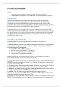
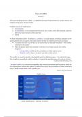
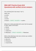
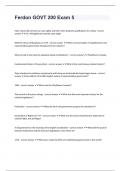
Tabel 3.1: Foto’s met UV licht en Groen licht van HeLa cellen behandeld met verschillende
Drugs: Vinblastine en Cytochalasin D. Ook een test met 0.05 mM concentratie CD.
Bij de controle is er geen Drug.
Glaasje UV licht Groen licht Merged
1 Cytochalasin
D
2 Controle
3 Vinblastine
4 0.05mM
Doel
- Het opzetten van een goed protocol om HeLa cellen aan te kleuren.
- Onderzoeken wat het effect is van Vinblastine en Cytochalasine D is op cellen.
Hypothese
Door het toevoegen van hoge concentratie vinblastine worden microtubili en
intermediaire filamenten gedepolymeriseerd(J.H. Temmink et all., 1981) . Omdat
vinblastine niet op de actinefilamenten werkt verwachten we hier intacte actine
filamenten. Dit is met de microscoop goed te zien omdat de labeloplossing op de actine
filamenten kan hechten en een fluorescente kleur kan geven.
Cytochalasine D zorg voor depolarisatie van actine filamenten (K. Mortensen et all.,
2003). Hierdoor kan de labeloplossing niet binden aan actine en zal er niets te zien zijn
met de microscoop. In de controle (waar geen drug is toegevoegd) worden filamenten
niet afgebroken, hier kan labeloplossing binden aan actine en worden het cytoskelet
zichtbaar bij de microscoop.
Materiaal & Methode
Voor materialen zie Practicum handleiding Visualising Cells 5052CEO12Y.
1. Drug behandelen: Allereerst is de drug behandeling van Cytochalasin D en Vinblastine
uitgevoerd op 4 dekglaasjes met HeLa cellen. Aan de HeLa cellen zijn verschillende
hoeveelheden vloeistof toegevoegd:
1- 1L DMSO + 999 L PBS (controle)
2- 1L van 9,8 mM Cytocalasin D + 999 L PBS
3- 1 L van 5,5 mM Vinblastine + 999 L PBS
4- concentratie van 0.05mM Cytocalasin D
De incubatietijd voor Cytochalasin D is 30 minuten, voor vinblastine, controle en
0.05mM Cytocalasin D was de incubatietijd 45 minuten.
2. Fixeren: Vervolgens zijn de cellen 15 minuten gefixeerd met 100 L PBS/FA. De fixatie
is gestopt door het toevoegen van 100 L 0.1 M Tris-HCl. Na fixatie zijn de HeLa cellen
3x gewassen met 100 L PBS.
3. Permeabel maken: Daarna permeabel gemaakt middels 10 min 100L PBS-Tx100. Na
het permeabel maken zijn de cellen opnieuw 3x gewassen met 100 L PBS.
4. Blokkeren: Vervolgens zijn de hydrofobe structuren afgedekt om aspecifieke kleuring
te voorkomen middels 100L BSA/PBS voor 20 minuten.
5. Kleuring: Voor het maken van de kleuring is gebruik gemaakt van een
labelingsoplossing: 50 L 0.1M Rhodamine-phalloidin+DAPI in PBS voor 20 minuten.
Na kleuring opnieuw 3x wassen met 100 L PBS
6 Insluiten: Tot slot zijn de HeLa cellen op het preparaat gereed gemaakt de microscoop.
Hierbij. is gebruik gemaakt van een druppeltje Mowiol (insluiten). De dekglaasjes met
, HeLa cellen zijn vervolgens omgekeerd op het druppeltje Mowiol gelegd en vastgezet
met nagellak. Onder de microscoop is het cytoskelet bekenen.
Voor het bewerken van de foto’s is ImageJ gebruikt. Van elk beeld zijn twee foto’s
gemaakt; 1 met UV licht en 1 met groen licht. Vervolgens zijn met de afbeeldingen
scherper gemaakt met Smooth. Als er ruis aanwezig was op de foto’s zijn deze
weggewerkt met Smooth filter (Process Smooth). Daarna is hier Merge Channels op
uitgevoerd door (image Color Merge channels). Hierbij is er voor gekozen dat de
kernen rood gekleurd werden en actine filamenten groen. Dit omdat hierbij het verschil
het duidelijkst was.
Resultaten
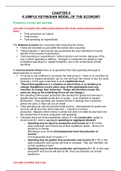
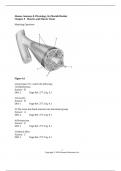
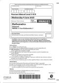
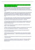
Tabel 3.1: Foto’s met UV licht en Groen licht van HeLa cellen behandeld met verschillende
Drugs: Vinblastine en Cytochalasin D. Ook een test met 0.05 mM concentratie CD.
Bij de controle is er geen Drug.
Glaasje UV licht Groen licht Merged
1 Cytochalasin
D
2 Controle
3 Vinblastine
4 0.05mM