Vascular surgery
Chronic Limb Ischaemia and Peripheral Vascular Disease
Peripheral vascular disease
Stenotic +/- atherosclerotic disease of peripheral arteries producing symptoms + signs of ischaemia
Peripheral infra-renal aorta to the feet
Common in elderly population but only 1-2% develop critical limb ischaemia
Mild: intermittent claudication, pain relieved by rest
Mod: short distance claudication < 100m
Severe: v. short distance claudication, nocturnal pain
Rest pain, ulceration/tissue loss critical
limb ischaemia
History: Typical CV risk factors, associated with IHD,
ischaemic stroke
- Intermittent claudication
o Claudication distance is the same
every time
o Worse up hill or walking fast
o Relieved by rest
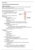
o Calf, thigh, buttock muscle groups affected depending on level of PVD
o Unilateral/bilateral symptoms – one limb almost always worse than other
- Red flag symptoms
o Very short distance claudication < 50m
o Nocturnal foot pain
Neuro-ischaemic pain ( BP at night hydrostatic pressure)
Patient awoken by pain – needs to hang foot out of bed/get up and walk around
o Pain at rest (in foot) – typically requires opiate analgesia
o Ulceration or tissue loss
Examination
- Appearance
o Dry, shiny, thin skin
o Loss of hair
o Loss of muscle bulk
o Thickened nails
o Ulcers – toe and pressure areas
o Necrosis/tissue loss
- Pulses: femoral, popliteal, dorsalis pedis, posterior tibial
o Palpable foot pulses? Not significant PVD
o Beware of the easily palpable popliteal pulse ?popliteal artery aneurysm
- Handheld Doppler
o Monophasic signal PVD
- Ankle Brachial Pressure Index
o > 1.2 Upper limb PVD or incompressible vessles e.g. infra-popliteal Ca2+ in DM
o ? Absolute pressure
o < 0.9 PVD
o ABPI < 0.3 or absolute pressure < 40mmHg ~ critical limb ischaemia
- Buerger’s sign (a) – significant lower limb ischaemia – cold, red foot
o Elevating the foot results in significant pallor and venous guttering
, o Lowering the foot results in dependent rubor ‘redness’
o Foot becomes cyanotic ‘sunset’ foot
- Arterial ulcers (b)
o ‘Punched out’
o Occur at toes or pressure points
o Painful
o Associated with severe PVD and signs
- Necrosis/tissue loss threatened limb (c)
Differentials
1. Arthritis
a. Joint stiffness in am + symptoms worse at end of day
b. Good days and bad days
2. Spinal stenosis
a. Relieved by sitting down/leaning forwards
3. Sciatica/lumbar spine radiculopathy
a. Paraesthesia at toes in dermatomal distribution
b. Posterior thigh symptoms – not quads
c. Symptoms at rest/in bed/positional
d. Only proximal symptoms
If patient gets pain at rest it isn’t claudication
Management
1. Best medical therapy
a. Stop smoking
b. Anti-platelet (aspirin/clopidogrel)
c. ? Aspirin + low dose rivaroxaban
d. Statin
e. Diabetic glycaemic control
f. BP control
g. Exercise – 30 mins walking x3/week
Investigations
- Arterial duplex
o Non-invasive but time consuming and not available OOH
o Operator dependent
o Calcification obscures view esp in infra-popliteal disease
o Poor views of distal aorta/iliac arteries due to bowel gas
- MR angiogram – gives view of lumen of vessels but not much information about walls
o Not available OOH, contraindications with metalwork
, o Patients with eGFR < 30 may develop nephrogenic systemic fibrosis (gadolinium contrast)
which may be fatal
- CT angiogram
o Available OOH, may result in contrast induced nephropathy
o Calcification may make imaging more difficult
- Catheter angiogram – ‘gold standard’ but invasive
o Digital subtraction angiogram – only artery can be seen
Infra-popliteal disease
Typically affects diabetics – develop severe PVD and calcification below the knee
- Often co-existing peripheral neuropathy – unnoticed trauma
- Ulceration complicated by infection
- May not have claudication but may present with tissue loss/ulceration +/- infection
- O/E
o Femoral + popliteal pulses but no PT/DP
o Unreliable ABPI due to incompressible vessels from calcium
Treatment
- Angioplasty or distal bypass
o Fem-distal bypass
Bypass between common femoral a. and one of the 3 below knee arteries using
great saphenous vein
Typically ipsilateral GSV in reversed configuration
Non-reversed/insitu GSV can be used but valves must be removed
If insufficient GSV can take contralateral GSV/upper limb vein and splice the vein
Prosthetic bypass can be performed using dacron/PTFE +/- vein cuff
- Debridement or drainage of infection
Femoral-popliteal disease
Superficial femoral a./popliteal a.
- Calf claudication
- Claudication critical limb ischaemia
- Femoral pulse only, no popliteal pulse or distal pedal pulses
Treatment
- Angioplasty +/- stent
- Fem-pop bypass
Chronic Limb Ischaemia and Peripheral Vascular Disease
Peripheral vascular disease
Stenotic +/- atherosclerotic disease of peripheral arteries producing symptoms + signs of ischaemia
Peripheral infra-renal aorta to the feet
Common in elderly population but only 1-2% develop critical limb ischaemia
Mild: intermittent claudication, pain relieved by rest
Mod: short distance claudication < 100m
Severe: v. short distance claudication, nocturnal pain
Rest pain, ulceration/tissue loss critical
limb ischaemia
History: Typical CV risk factors, associated with IHD,
ischaemic stroke
- Intermittent claudication
o Claudication distance is the same
every time
o Worse up hill or walking fast
o Relieved by rest
o Calf, thigh, buttock muscle groups affected depending on level of PVD
o Unilateral/bilateral symptoms – one limb almost always worse than other
- Red flag symptoms
o Very short distance claudication < 50m
o Nocturnal foot pain
Neuro-ischaemic pain ( BP at night hydrostatic pressure)
Patient awoken by pain – needs to hang foot out of bed/get up and walk around
o Pain at rest (in foot) – typically requires opiate analgesia
o Ulceration or tissue loss
Examination
- Appearance
o Dry, shiny, thin skin
o Loss of hair
o Loss of muscle bulk
o Thickened nails
o Ulcers – toe and pressure areas
o Necrosis/tissue loss
- Pulses: femoral, popliteal, dorsalis pedis, posterior tibial
o Palpable foot pulses? Not significant PVD
o Beware of the easily palpable popliteal pulse ?popliteal artery aneurysm
- Handheld Doppler
o Monophasic signal PVD
- Ankle Brachial Pressure Index
o > 1.2 Upper limb PVD or incompressible vessles e.g. infra-popliteal Ca2+ in DM
o ? Absolute pressure
o < 0.9 PVD
o ABPI < 0.3 or absolute pressure < 40mmHg ~ critical limb ischaemia
- Buerger’s sign (a) – significant lower limb ischaemia – cold, red foot
o Elevating the foot results in significant pallor and venous guttering
, o Lowering the foot results in dependent rubor ‘redness’
o Foot becomes cyanotic ‘sunset’ foot
- Arterial ulcers (b)
o ‘Punched out’
o Occur at toes or pressure points
o Painful
o Associated with severe PVD and signs
- Necrosis/tissue loss threatened limb (c)
Differentials
1. Arthritis
a. Joint stiffness in am + symptoms worse at end of day
b. Good days and bad days
2. Spinal stenosis
a. Relieved by sitting down/leaning forwards
3. Sciatica/lumbar spine radiculopathy
a. Paraesthesia at toes in dermatomal distribution
b. Posterior thigh symptoms – not quads
c. Symptoms at rest/in bed/positional
d. Only proximal symptoms
If patient gets pain at rest it isn’t claudication
Management
1. Best medical therapy
a. Stop smoking
b. Anti-platelet (aspirin/clopidogrel)
c. ? Aspirin + low dose rivaroxaban
d. Statin
e. Diabetic glycaemic control
f. BP control
g. Exercise – 30 mins walking x3/week
Investigations
- Arterial duplex
o Non-invasive but time consuming and not available OOH
o Operator dependent
o Calcification obscures view esp in infra-popliteal disease
o Poor views of distal aorta/iliac arteries due to bowel gas
- MR angiogram – gives view of lumen of vessels but not much information about walls
o Not available OOH, contraindications with metalwork
, o Patients with eGFR < 30 may develop nephrogenic systemic fibrosis (gadolinium contrast)
which may be fatal
- CT angiogram
o Available OOH, may result in contrast induced nephropathy
o Calcification may make imaging more difficult
- Catheter angiogram – ‘gold standard’ but invasive
o Digital subtraction angiogram – only artery can be seen
Infra-popliteal disease
Typically affects diabetics – develop severe PVD and calcification below the knee
- Often co-existing peripheral neuropathy – unnoticed trauma
- Ulceration complicated by infection
- May not have claudication but may present with tissue loss/ulceration +/- infection
- O/E
o Femoral + popliteal pulses but no PT/DP
o Unreliable ABPI due to incompressible vessels from calcium
Treatment
- Angioplasty or distal bypass
o Fem-distal bypass
Bypass between common femoral a. and one of the 3 below knee arteries using
great saphenous vein
Typically ipsilateral GSV in reversed configuration
Non-reversed/insitu GSV can be used but valves must be removed
If insufficient GSV can take contralateral GSV/upper limb vein and splice the vein
Prosthetic bypass can be performed using dacron/PTFE +/- vein cuff
- Debridement or drainage of infection
Femoral-popliteal disease
Superficial femoral a./popliteal a.
- Calf claudication
- Claudication critical limb ischaemia
- Femoral pulse only, no popliteal pulse or distal pedal pulses
Treatment
- Angioplasty +/- stent
- Fem-pop bypass