ECG Pictures Latest 2023 Graded A
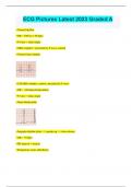
ECG Pictures Latest 2023 Graded A • Normal rhythm • HR = 300/4.4 = 68 bpm • P wave = sinus origin • QRS complex = preceded by P wave, normal • Normal sinus rhythm • Tall QRS complex, narrow, preceded by P wave • HR = <60 bpm (bradycardia) • P wave = sinus origin • Sinus bradycardia • Irregular rhythm (slow --> speeds up --> slows down) • HR = 70 bpm • PR interval = normal • Respiratory sinus arrhythmia • 2 sinus pauses (arrests) • P wave = sinus node origin • PR interval = close to being long --> 1st heart/AV block • Normal sinus rhythm, but with 2 sinus arrests • Rapid HR = 300/2.6 = 110 bpm • Normal rhythm • P wave = sinus node origin • PR interval = normal • QRS complex = normal, preceded by P wave • Sinus tachycardia o If you can't see a P wave --> supraventricular tachycardia • F waves present • Saw-tooth pattern • Associated with atrial valve disease that results in enlarged atria • Atrial flutter • Irregular rhythm • Right on border of ST segment depression (upsloping so okay) • Sinus tachycardia, with 2 PACs (Premature atrial contractions) o P-wave occurred during T-wave after QRS complex, altering ST segment = PAC • P-wave = none • Irregular rhythm and rate (fast) • QRS complex = narrow, normal • Supraventricular tachycardia • Irregularly irregular = no pattern + irregular • Irregular rhythm • HR = 90 bpm • Atrial fibrillation --> key is chaotic electrical baseline (bunch of P-waves) o Typically associated with enlarged atria • Regular rhythm • P-waves = changing shapes, and if you see at least 3 different shapes --> wandering atrial pacemaker o Occurs in at least 3 different areas of the atria • ST segment depression = myocardial ischemia o Elevation = heart attack • Supraventricular tachycardia • Short PR interval in 2 areas --> P wave affecting T-wave = PAC • Normal sinus rhythm, with 2 PACs • Paroxysmal = burst • Paroxysmal supraventricular tachycardia • Early beat followed by pause • QRS complex = narrow • Premature junctional complex (PJC) • Normal sinus rhythm with 1 PJC • Junctional rhythm --> no P-wave + narrow QRS + ST segment elevation o Multiple junctional escape beats in a row • HR = 40-60 bpm • 2 wide but different shaped QRS --> different origins • Fast rate • Multifocal PVCs --> from different site • Sinus tachycardia with 2 PVCs • Normal sinus rhythm --> ventricular tachycardia (monomorphic) • Wide QRS, preceded by P-wave • Slow conduction to ventricles --> bundle branch block • Intraventricular conduction block delay • PVC every other beat --> Bigemini • Ventricular fibrillation --> requires defibrillation as soon as possible • Associated with CAD • Polymorphic ventricular tachycardia --> torsade de Puente • HR = ~40 bpm • P waves = none • QRS complex = little wide • Accelerated idioventricular rhythm • Long PR interval = 1st degree AV block • HR = ~50 bpm • Sinus bradycardia with 1st degree AV block • Extra P-wave --> dropped QRS • Second degree AV block type 1 (PR interval gets longer then stops = P wave not leading to QRS) o Wenkiback • Bradycardia • 3rd degree AV block o Clue is that PR interval keeps changing o Means the P-waves are not related to QRS • P-waves after QRS • AV junction is where activity starts o Junctional rhythm (40-60 bpm) o Accelerated junctional rhythm (>60 bpm) • Wide QRS + rhythm = idioventricular rhythm (20-40 bpm) o HR = ~25 bpm • HR < 20 = agonal rhythm • Supraventricular tachycardia (no P-wave + narrow QRS) before hump • Becomes sinus tachycardia after hump • 2 uni-focal PVCs with normal rhythm in between • Normal sinus rhythm with 2 uni-focal PVCs • F-waves • Saw-tooth pattern • Atrial flutter
Written for
- Institution
- ECG Pictures
- Course
- ECG Pictures
Document information
- Uploaded on
- October 31, 2023
- Number of pages
- 10
- Written in
- 2023/2024
- Type
- Exam (elaborations)
- Contains
- Questions & answers
Subjects
-
ecg pictures latest 2023 graded a
Also available in package deal