Ophthalmology Note Cards Latest Update 2023
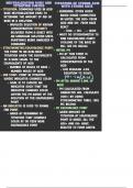
Ophthalmology Note Cards Latest Update 2023 Viral conjunctivitis - >>-characterized by watery discharge from the eye and a swollen pre-auricular lymph node -tends to be unilateral before hitting the other eye. The onset occurs within 1-3 days. -Subepithelial infiltrates are scarring of the cornea due to irritation from dead viral particles -Adenoviruses are extremely contagious, so all exams should be done with gloves and Q tips. -There are NO TREATMENTS, so just avoid spreading it with fastidious hand washing. Bacterial conjunctivitis - >>-more insidious onset than viral -purulent discharge that's sticky and crusty -treated with a topical antibiotic, usually a gel. If it's N. gonorrhea, give IM ceftriaxone. -In children, we're particularly worried about N. gonorrheae and H. influenza Corneal ulcer - >>-white area on the eye with thinning and ulceration -from bacteria or fungi -most common cause is from a contact lens, though trauma and other irritants can cause it too --> cells infiltrate the stroma, and some WBCs can accumulate in the anterior chamber. -TX: anesthetize, scrape, and culture the eye, because the rare side effects are dangerous Herpetic corneal ulcer - >>-lesion on the cornea with a dendritic form -no history of trauma -Fluorescein stains the lesion -Pain, photophobia, redness, and a foreign body sensation -TX: just like any other HSV infection, giving acyclovir helps. In this case, it prevents inflammation. Cycloplegic drops can freeze the ciliary muscles and prevent spasm from accommodation. Corneal abrasion - >>-assessed using fluorescein to see if there's a foreign body or just the scrape. -pain and photophobia. TX: Prophylactic antibiotics and cycloplegic drops are given. -If there's actually a foreign body, there's an anterior chamber reaction. The Tx. is the same. --> The eyelids should be everted to check for a conjunctival foreign body. In that case, there are lines up and down the cornea due to scraping when blinking. Corneoscleral lacerations - >>-usually a history of penetrating trauma -pai, redness, and loss of vision -Tx: tetanus toxoid and IV antibiotics; surgery is done to repair it. -Intraocular foreign bodies are usually from penetrating missiles. The tx. is the same. -->The loss of vision isn't always there. It depends on where within the eye it lands. Vitreous hemorrhage - >>-involves a SUDDEN DROP IN ACUITY with a bunch of floaters appearing in visual fields -Key thing to rule out: retinal tear or choroidal rupture. Major risks are DIABETES and SICKLE CELL. -These patients need no therapy. Keep the head elevated and avoid aspirin and the Valsalva. Retinal detachment - >>-presents similarly to vitreous hemorrhage, with flashing lights and floaters. A "CURTAIN" is pathognomonic. -occurs more often in myopic people due to retinal thinning. As such, check both eyes. -Tears look like horseshoes (that's more from trauma). If it's from diabetes, there's just pulling. Subconjunctival hemorrhage - >>-totally BENIGN, just a patch of blood. Work it up if it becomes recurrent. Dry eye syndrome - >>-causes a foreign body sensation that's worsened by breezes (even air conditioning). -these patients have decreased tear break-up time and a scant tear meniscus -artificial tears are too watery to be used regularly. Topical steroids and cyclosporine are helpful. -Plugs can be put in the lacrimal duct to make the tears stay in the eye longer. Blepharitis - >>-characterized by a collection of crusting (cells, bacteria, and oils) on the eyelids. -It's associated with ROSACEA. -Tx: warm compresses, lid hygiene, and artificial tears. Oral doxycycline helps by thinning the secretions of the oils (it's an acne tx). Chalazion - >>-appears as a bump on an eyelid due to a blocked Meibomian gland -It's just lipogranulomatous inflammation, NOT an infection. It's associated with blepharitis. Tears - >>-lubricate and protect the cornea, as they contain immunoglobulins, lysozymes, and lactoferrin Tear film - >>-provides O2 and nutrients for the ocular surface. -keeps vision clear Tears: Aqueous layer - >>-created by lacrimal glands -dysfunctional in Sjogren syndrome Tears: lipid layer - >>-created by Meibomian glands -Ocular rosacea and blepharitis screw the glands up Tears: mucinous layer - >>-derived from conjunctival goblet cells, which can be destroyed in SJS Cornea - >>-"windshield for the lens" that provides 2/3 of refraction (lens is other 1/3) -completely avascular Corneal epithelium - >>-can be scratched or infected by HSV, in which can there are dendrites Bowman's layer of cornea - >> CONTINUES....
Written for
- Institution
- Ophthalmology
- Course
- Ophthalmology
Document information
- Uploaded on
- October 26, 2023
- Number of pages
- 24
- Written in
- 2023/2024
- Type
- Class notes
- Professor(s)
- Proff
- Contains
- All classes
Subjects
- ophthalmology
- viral conjunctivitis
- corneal ulcer
- herpetic corneal ulcer
-
ophthalmology note cards latest update 2023
-
bacterial conjunctivitis
Also available in package deal