CYTOSKELETON
− the cytoplasm contains an array of fibrous proteins collectively called the cytoskeleton
− 3 classes of fibers compose the cytoskeleton: microfilaments (MF) → built of actin protein,
microtubules (MT) → built of tubulin protein polymers, intermediate filaments (IF)
− dynamic network of long polymeric filaments → all these fibers are long chains of multiple copies of
one or more small protein subunits
− their assembly and break up are regulated by a large variety of cytoskeleton-binding proteins: actin
and MTs
− function:
o intracellular organization
o cellular morphology
o cellular movement: muscle contraction, cell migration, intracellular transport, movement,
mitosis) → actin & MTs
o multicellular organization, regulation of gene expression → actin & IFs
MICROFILAMENT
1. molecular actin filament
organization/dynamics
− G-actin (globular) = monomer of 4
subunits with an ATP-binding cleft where
Mg2+ sits, intrinsically polar structure
− F-actin (filament) = due to their polarity,
G-actins assemble into a repeating unit
− ATPase activity is activated in F-actin and
causes polarity (asymmetry) of actin
filaments
− ATP/ADP binding regulates actin
conformation (not needed for energy)
− G-actin will be incorporated into F-actin
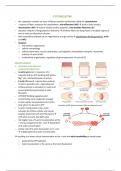
only when it is bound to ATP Examples of MF-based structures (MFs are depicted in red)
− G-actin incorporation into F-actin
activates its ATPase activity and causes
polarity (asymmetry of actin filaments) →
ATP-actin converts into ADP-actin
− 10x higher rate of G-actin incorporation at the
+ end as compared to the - end → dependent
of G-actin concentration
− similar rate of G-actin dissociation at +/- end
→ independent of G-actin concentration
→ resulting in a lower critical concentration at the + end and actin treadmilling at steady state:
o powered by ATP hydrolysis
o total incorporation is the same as the total dissociation
1
, o net elongation at + end and shortening at – end
o basis for actin dynamics
o caused by net incorporation/dissociation of G-actin-ATP/ADP at the +/− end
− cellular actin conc. is high → without regulation high spontaneous polymerisation at both ends
− capping proteins:
o stabilize F-actin
o CapZ inhibits incorporation at the + end
o tropomodulin inhibits dissociation at the –
end
o found in skeletal muscle where actin is very
stable
− F-actin turnover:
o assembly rate improved
o profilin = attaches to actin and promotes ATP
loading onto it → more ATP actin is made, binds
at the + end of F-actin thus preventing G-actin-
ATP incorporation at the − end, stimulator of
actin assembly
o thymosin-β4 = binds and sequesters G-actin-ATP
and thus lowers its free concentration, competes
with profilin for ATP-actin binding
o cofilin = binds to ADP-actin and severs F-actin
filaments, destabilizes ADP-bound F-actin and
promotes turnover/treadmilling
o gelsolin = F-actin severing
2. mechanisms of filament assembly
− nucleation factors stimulate formation of new actin filaments
− nucleation = G-actin clusters around nucleus
− formin:
o stimulates nucleation → dimer of formin FH2 domain binds profilin-ATP-G-actin complex
o stimulates formation of (long) actin stress fibers
o remains bound to the + end → prevents binding of capping proteins
o activated via the activation of the small GTPase Rho (Ras’s homology)
− regulation of Rho family (Rho, Rac, Cdc42):
o GEF = guanine exchange factor
o GAP = GTPase activating protein
2
, o GDI = GDP-dissociation inhibitor – prevents activation and recruitment to the plasma
membrane
− nucleation of linear actin filaments by Rho-dependent activation of formin → RBD = Rho binding
domain
− ARP2/3 (actin-related protein)
actin nucleation complex:
o forms branched actin
filaments
o NPFs = nucleation-
promoting factors,
WASp/WAVE, RBD
domain → activated
by active Rac/Cdc42-
GTP or by PIP2
o binding of
WASp/WAVE to an
actin monomer and ARP2/3
o binding of ARP2/3 (with
already bound WASp/WAVE
and actin) to the side of F-
actin chain
o elongation of actin branch
o ARP2/3 remains associated
with the – end
− phagocytosis is driven by actin
polymerization
− Listeria bacteria → ARP2/3 complex
3
− the cytoplasm contains an array of fibrous proteins collectively called the cytoskeleton
− 3 classes of fibers compose the cytoskeleton: microfilaments (MF) → built of actin protein,
microtubules (MT) → built of tubulin protein polymers, intermediate filaments (IF)
− dynamic network of long polymeric filaments → all these fibers are long chains of multiple copies of
one or more small protein subunits
− their assembly and break up are regulated by a large variety of cytoskeleton-binding proteins: actin
and MTs
− function:
o intracellular organization
o cellular morphology
o cellular movement: muscle contraction, cell migration, intracellular transport, movement,
mitosis) → actin & MTs
o multicellular organization, regulation of gene expression → actin & IFs
MICROFILAMENT
1. molecular actin filament
organization/dynamics
− G-actin (globular) = monomer of 4
subunits with an ATP-binding cleft where
Mg2+ sits, intrinsically polar structure
− F-actin (filament) = due to their polarity,
G-actins assemble into a repeating unit
− ATPase activity is activated in F-actin and
causes polarity (asymmetry) of actin
filaments
− ATP/ADP binding regulates actin
conformation (not needed for energy)
− G-actin will be incorporated into F-actin
only when it is bound to ATP Examples of MF-based structures (MFs are depicted in red)
− G-actin incorporation into F-actin
activates its ATPase activity and causes
polarity (asymmetry of actin filaments) →
ATP-actin converts into ADP-actin
− 10x higher rate of G-actin incorporation at the
+ end as compared to the - end → dependent
of G-actin concentration
− similar rate of G-actin dissociation at +/- end
→ independent of G-actin concentration
→ resulting in a lower critical concentration at the + end and actin treadmilling at steady state:
o powered by ATP hydrolysis
o total incorporation is the same as the total dissociation
1
, o net elongation at + end and shortening at – end
o basis for actin dynamics
o caused by net incorporation/dissociation of G-actin-ATP/ADP at the +/− end
− cellular actin conc. is high → without regulation high spontaneous polymerisation at both ends
− capping proteins:
o stabilize F-actin
o CapZ inhibits incorporation at the + end
o tropomodulin inhibits dissociation at the –
end
o found in skeletal muscle where actin is very
stable
− F-actin turnover:
o assembly rate improved
o profilin = attaches to actin and promotes ATP
loading onto it → more ATP actin is made, binds
at the + end of F-actin thus preventing G-actin-
ATP incorporation at the − end, stimulator of
actin assembly
o thymosin-β4 = binds and sequesters G-actin-ATP
and thus lowers its free concentration, competes
with profilin for ATP-actin binding
o cofilin = binds to ADP-actin and severs F-actin
filaments, destabilizes ADP-bound F-actin and
promotes turnover/treadmilling
o gelsolin = F-actin severing
2. mechanisms of filament assembly
− nucleation factors stimulate formation of new actin filaments
− nucleation = G-actin clusters around nucleus
− formin:
o stimulates nucleation → dimer of formin FH2 domain binds profilin-ATP-G-actin complex
o stimulates formation of (long) actin stress fibers
o remains bound to the + end → prevents binding of capping proteins
o activated via the activation of the small GTPase Rho (Ras’s homology)
− regulation of Rho family (Rho, Rac, Cdc42):
o GEF = guanine exchange factor
o GAP = GTPase activating protein
2
, o GDI = GDP-dissociation inhibitor – prevents activation and recruitment to the plasma
membrane
− nucleation of linear actin filaments by Rho-dependent activation of formin → RBD = Rho binding
domain
− ARP2/3 (actin-related protein)
actin nucleation complex:
o forms branched actin
filaments
o NPFs = nucleation-
promoting factors,
WASp/WAVE, RBD
domain → activated
by active Rac/Cdc42-
GTP or by PIP2
o binding of
WASp/WAVE to an
actin monomer and ARP2/3
o binding of ARP2/3 (with
already bound WASp/WAVE
and actin) to the side of F-
actin chain
o elongation of actin branch
o ARP2/3 remains associated
with the – end
− phagocytosis is driven by actin
polymerization
− Listeria bacteria → ARP2/3 complex
3