Lecture 23 Development of the brain (part 1)
All behavioral development depends on the extent of brain
development. Brain development is dependent upon both
experience and genetics. The brain has a great deal of plasticity and
can recover over time. The brain is not a single system. Different
parts of the brain control different functions. Brain cells are nerve
(neurons) and glial cells. Information from outside the body is stored
into neurons and their communication to one another (‘network’).
The reaction to this information requires networks of communicating
neurons (not necessarily in the same brain part). Neuronal
communication is largely influenced by glial cells ‘enwrapping’
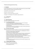
neurons (against the Neuron-Dogma). At 25 days, the neural tube
has developed and the first two branchial arches, which will form the
face and the viserocranium. By folding of the neural tube, the
different parts of the brain will be formed. The brain is comprised of three main areas:
1. Hind brain (rhombencephalon)
- Cerebellum and pons (metencephalon)
- Medulla oblongata (myelencephalon)
2. Midbrain (mesencephalon)
- Tectum and tegmentum (substantial
nigra)
3. Forebrain (prosencephalon)
- Diencephalon
- Telencephalon (cerebrum/cortex)
Growth of the brain occurs from the inside out and the bottom up. Children are born with 100 billion
(0.1 x 10¹²) neurons. There are around 1000 synaptic connections for each cell.
The human central nervous system begins to form when the embryo is about two weeks old. The dorsal
surface thickens and forms a neural tube surrounding a fluid filled cavity. The forward end enlarges and
differentiates into the hindbrain, midbrain and forebrain. The rest of the neural tube becomes the spinal
cord. In week 2-4 the primitive streak forms. The mesoderm was made, especially the notochord, which
was the inducer of the ectoderm (were made from epiblast). This formed the neural plate, which would
become the neural groove and, in the end, the neural tube. The structural components of the adult
brain are the nuclei (clusters of neuronal cell bodies), tracts (connecting pathways between different
parts of the brain) and the cortex (a dens rind of neuronal cell bodies covering fore-, mid- and hindbrain
structures. The functional component are the neuronal systems, which are collections of interconnected
nuclei that serve a common function (e.g., the visual system).
Cephalization is the gradual emergence of gyri and sulci (increasing surface of the brain). The walnut
shaped structures are called the cerebral hemispheres (left and right brain halve). The cerebral
hemispheres are kept together by the corpus callosum. The telencephalon is attached to the thalamus
with the internal capsule. The telencephalon is a thin layer of cells that covers both hemispheres. The
cortex consists of four parts:
1. Visual cortex high-level visual processing
, 2. Parietal cortex sensory integration and visual-motor processing
3. Temporal cortex auditory/visual processing and receptive language
4. Frontal cortex higher-level cognition, motor control and expressive language
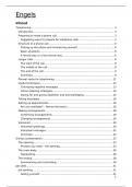
Key players of the nervous system are neurons. They
message transmitting cells. There are many subtypes
depending on the neurotransmitter they make and release
(chemical signal between neurons). The NT is released upon
an action potential (electrical activity within a neuron).
Multiple synaptic inputs have to happen at the same time to
generate an action potential. The synapse is the part where
the neurotransmitter is released. It is composed of a
presynaptic and a postsynaptic side. The type on message depends on the type of receptors. The classes
of neurons are dependent of: the number of neurites, the shape of the dendrites, the length of the
axon, the function, the transmitter secretion and if it is activating or inhibiting.
Glial cells, such as microglia, astrocytes and oligodendrocytes, are also key players. They are responsible
for maintaining neurons and communication. Oligodendrocytes produce myelin that surrounds the axon
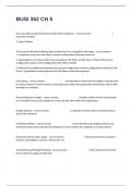
and increases the speed of the electrical transmission. There are 8 phases in embryonic and fetal
development at a cellular level. The 8 stages are sequential for a given neuron, but all are occurring
simultaneously throughout fetal development in different parts of the brain.
, Mitosis Death
Migration Rearrangement
Aggregation &
Myelination
differentiation
Synaptogenesis
The neuroepithelial layer gives rise to neuroblasts. In turn, neuroblasts give rise to neurons and
gliablasts. At early stages, a stem cell generates radial glial cells and neuroblasts. Later, it undergoes a
specific asymmetric division (the ‘switch point’) at which it changes from making neurons to making glia.
Radial glial cells act as guide wires for the migration of neurons. Migration of neurons happen with the
usage of growth cones. Growth cones (microfilaments and actin) crawl forward, dragging the extending
axons behind them. Their extension is controlled by cues in their outside environment that direct them
All behavioral development depends on the extent of brain
development. Brain development is dependent upon both
experience and genetics. The brain has a great deal of plasticity and
can recover over time. The brain is not a single system. Different
parts of the brain control different functions. Brain cells are nerve
(neurons) and glial cells. Information from outside the body is stored
into neurons and their communication to one another (‘network’).
The reaction to this information requires networks of communicating
neurons (not necessarily in the same brain part). Neuronal
communication is largely influenced by glial cells ‘enwrapping’
neurons (against the Neuron-Dogma). At 25 days, the neural tube
has developed and the first two branchial arches, which will form the
face and the viserocranium. By folding of the neural tube, the
different parts of the brain will be formed. The brain is comprised of three main areas:
1. Hind brain (rhombencephalon)
- Cerebellum and pons (metencephalon)
- Medulla oblongata (myelencephalon)
2. Midbrain (mesencephalon)
- Tectum and tegmentum (substantial
nigra)
3. Forebrain (prosencephalon)
- Diencephalon
- Telencephalon (cerebrum/cortex)
Growth of the brain occurs from the inside out and the bottom up. Children are born with 100 billion
(0.1 x 10¹²) neurons. There are around 1000 synaptic connections for each cell.
The human central nervous system begins to form when the embryo is about two weeks old. The dorsal
surface thickens and forms a neural tube surrounding a fluid filled cavity. The forward end enlarges and
differentiates into the hindbrain, midbrain and forebrain. The rest of the neural tube becomes the spinal
cord. In week 2-4 the primitive streak forms. The mesoderm was made, especially the notochord, which
was the inducer of the ectoderm (were made from epiblast). This formed the neural plate, which would
become the neural groove and, in the end, the neural tube. The structural components of the adult
brain are the nuclei (clusters of neuronal cell bodies), tracts (connecting pathways between different
parts of the brain) and the cortex (a dens rind of neuronal cell bodies covering fore-, mid- and hindbrain
structures. The functional component are the neuronal systems, which are collections of interconnected
nuclei that serve a common function (e.g., the visual system).
Cephalization is the gradual emergence of gyri and sulci (increasing surface of the brain). The walnut
shaped structures are called the cerebral hemispheres (left and right brain halve). The cerebral
hemispheres are kept together by the corpus callosum. The telencephalon is attached to the thalamus
with the internal capsule. The telencephalon is a thin layer of cells that covers both hemispheres. The
cortex consists of four parts:
1. Visual cortex high-level visual processing
, 2. Parietal cortex sensory integration and visual-motor processing
3. Temporal cortex auditory/visual processing and receptive language
4. Frontal cortex higher-level cognition, motor control and expressive language
Key players of the nervous system are neurons. They
message transmitting cells. There are many subtypes
depending on the neurotransmitter they make and release
(chemical signal between neurons). The NT is released upon
an action potential (electrical activity within a neuron).
Multiple synaptic inputs have to happen at the same time to
generate an action potential. The synapse is the part where
the neurotransmitter is released. It is composed of a
presynaptic and a postsynaptic side. The type on message depends on the type of receptors. The classes
of neurons are dependent of: the number of neurites, the shape of the dendrites, the length of the
axon, the function, the transmitter secretion and if it is activating or inhibiting.
Glial cells, such as microglia, astrocytes and oligodendrocytes, are also key players. They are responsible
for maintaining neurons and communication. Oligodendrocytes produce myelin that surrounds the axon
and increases the speed of the electrical transmission. There are 8 phases in embryonic and fetal
development at a cellular level. The 8 stages are sequential for a given neuron, but all are occurring
simultaneously throughout fetal development in different parts of the brain.
, Mitosis Death
Migration Rearrangement
Aggregation &
Myelination
differentiation
Synaptogenesis
The neuroepithelial layer gives rise to neuroblasts. In turn, neuroblasts give rise to neurons and
gliablasts. At early stages, a stem cell generates radial glial cells and neuroblasts. Later, it undergoes a
specific asymmetric division (the ‘switch point’) at which it changes from making neurons to making glia.
Radial glial cells act as guide wires for the migration of neurons. Migration of neurons happen with the
usage of growth cones. Growth cones (microfilaments and actin) crawl forward, dragging the extending
axons behind them. Their extension is controlled by cues in their outside environment that direct them